

206
H E N R I E T T E HERLANT-MEEWIS
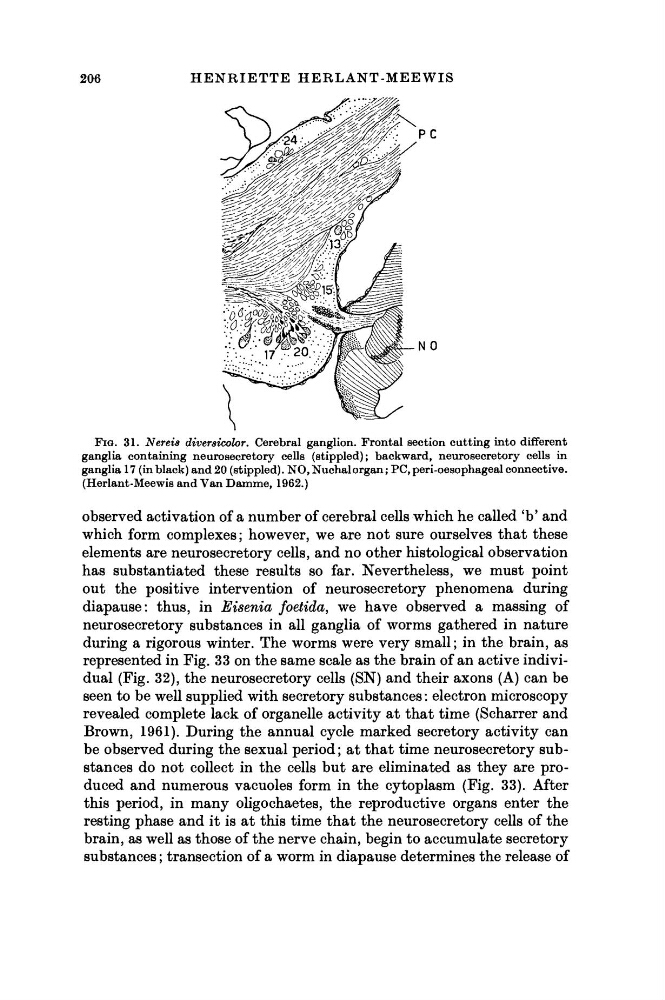
FIG. 31. Nereis diversicolor. Cerebral ganglion. Frontal section cutting into different
ganglia containing neurosecretory cells (stippled); backward, neurosecretory cells in
ganglia 17 (in black) and 20 (stippled). NO, Nuchal organ ; PC, peri-oesophageal connective.
(Herlant-Meewis and Van Damme, 1962.)
observed activation of a number of cerebral cells which he called 'b' and
which form complexes ; however, we are not sure ourselves that these
elements are neurosecretory cells, and no other histological observation
has substantiated these results so far. Nevertheless, we must point
out the positive intervention of neurosecretory phenomena during
diapause: thus, in Eisenia foetida, we have observed a massing of
neurosecretory substances in all ganglia of worms gathered in nature
during a rigorous winter. The worms were very small; in the brain, as
represented in Fig. 33 on the same scale as the brain of an active individual (Fig. 32), the neurosecretory cells (SN) and their axons (A) can be
seen to be well supplied with secretory substances : electron microscopy
revealed complete lack of organelle activity at that time (Scharrer and
Brown, 1961). During the annual cycle marked secretory activity can
be observed during the sexual period ; at that time neurosecretory substances do not collect in the cells but are eliminated as they are produced and numerous vacuoles form in the cytoplasm (Fig. 33). After
this period, in many oligochaetes, the reproductive organs enter the
resting phase and it is at this time that the neurosecretory cells of the
brain, as well as those of the nerve chain, begin to accumulate secretory
substances ; transection of a worm in diapause determines the release of
H E N R I E T T E HERLANT-MEEWIS
FIG. 31. Nereis diversicolor. Cerebral ganglion. Frontal section cutting into different
ganglia containing neurosecretory cells (stippled); backward, neurosecretory cells in
ganglia 17 (in black) and 20 (stippled). NO, Nuchal organ ; PC, peri-oesophageal connective.
(Herlant-Meewis and Van Damme, 1962.)
observed activation of a number of cerebral cells which he called 'b' and
which form complexes ; however, we are not sure ourselves that these
elements are neurosecretory cells, and no other histological observation
has substantiated these results so far. Nevertheless, we must point
out the positive intervention of neurosecretory phenomena during
diapause: thus, in Eisenia foetida, we have observed a massing of
neurosecretory substances in all ganglia of worms gathered in nature
during a rigorous winter. The worms were very small; in the brain, as
represented in Fig. 33 on the same scale as the brain of an active individual (Fig. 32), the neurosecretory cells (SN) and their axons (A) can be
seen to be well supplied with secretory substances : electron microscopy
revealed complete lack of organelle activity at that time (Scharrer and
Brown, 1961). During the annual cycle marked secretory activity can
be observed during the sexual period ; at that time neurosecretory substances do not collect in the cells but are eliminated as they are produced and numerous vacuoles form in the cytoplasm (Fig. 33). After
this period, in many oligochaetes, the reproductive organs enter the
resting phase and it is at this time that the neurosecretory cells of the
brain, as well as those of the nerve chain, begin to accumulate secretory
substances ; transection of a worm in diapause determines the release of