

REGENERATION IN ANNELIDS
205
neurosecretory cells of the ganglionic nuclei designated as S and Z become activated during the first hours of regeneration. (Herlant-Meewis
and Van Damme, 1962b) and Van Damme (1962), have described two
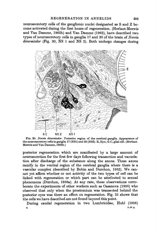
types of neurosecretory cells in ganglia 17 and 20 of the brain of Nereis
diversicolor (Fig. 30, NS 1 and NS 2). Both undergo changes during
GC
NS2
NS1
FIG. 30. Nereis diversicolor. Posterior region of the cerebral ganglia. Appearance of
the neurosecretory cells in ganglia 17 (NS1) and 20 (NS2). E, Eye ; G.C, glial cell. (HerlantMeewis and Van Damme, 1962b.)
posterior regeneration which are manifested by a large amount of
neurosecretion for the first few days following transection and vacuolation after discharge of the substance along the axons. These axons
ramify in the ventral region of the cerebral ganglia where there is a
vascular complex (described by Bobin and Durchon, 1952). We cannot yet affirm whether or not activity of the two types of cell can be
linked with regeneration or which part can be attributed to sexual
phenomena (Durchon, 1956a). At any rate, these observations corroborate the experiments of other workers such as Casanova (1955) who
observed that only when the prostomium was transected behind the
posterior eyes was there an effect on regeneration. Fig. 31 shows that
the cells we have described are not found beyond this point.
During caudal regeneration in two Lumbricidae, Hubl (1956)
G
A.M.4
205
neurosecretory cells of the ganglionic nuclei designated as S and Z become activated during the first hours of regeneration. (Herlant-Meewis
and Van Damme, 1962b) and Van Damme (1962), have described two
types of neurosecretory cells in ganglia 17 and 20 of the brain of Nereis
diversicolor (Fig. 30, NS 1 and NS 2). Both undergo changes during
GC
NS2
NS1
FIG. 30. Nereis diversicolor. Posterior region of the cerebral ganglia. Appearance of
the neurosecretory cells in ganglia 17 (NS1) and 20 (NS2). E, Eye ; G.C, glial cell. (HerlantMeewis and Van Damme, 1962b.)
posterior regeneration which are manifested by a large amount of
neurosecretion for the first few days following transection and vacuolation after discharge of the substance along the axons. These axons
ramify in the ventral region of the cerebral ganglia where there is a
vascular complex (described by Bobin and Durchon, 1952). We cannot yet affirm whether or not activity of the two types of cell can be
linked with regeneration or which part can be attributed to sexual
phenomena (Durchon, 1956a). At any rate, these observations corroborate the experiments of other workers such as Casanova (1955) who
observed that only when the prostomium was transected behind the
posterior eyes was there an effect on regeneration. Fig. 31 shows that
the cells we have described are not found beyond this point.
During caudal regeneration in two Lumbricidae, Hubl (1956)
G
A.M.4