

3 5 6
Z. R Y C H T E R
pletion, because the methods did not guarantee the invariable appearance of these defects in every embryo, a characteristic which is
necessary for serial investigations.
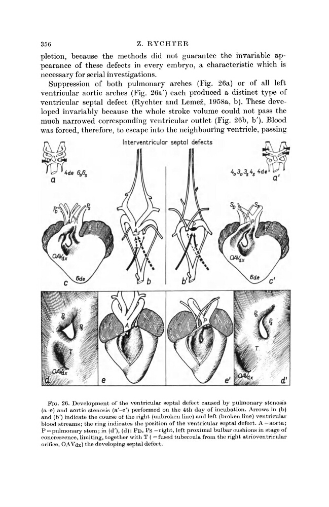
Suppression of both pulmonary arches (Fig. 26a) or of all left
ventricular aortic arches (Fig. 26a') each produced a distinct type of
ventricular septal defect (Rychter and Lemez, 1958a, b). These developed invariably because the whole stroke volume could not pass the
much narrowed corresponding ventricular outlet (Fig. 26b, b'). Blood
was forced, therefore, to escape into the neighbouring ventricle, passing
FIG. 26. Development of the ventricular septal defect caused by pulmonary stenosis
(a -e) and aortic stenosis (a'-e') performed on the 4th day of incubation. Arrows in (b)
and (b') indicate the course of the right (unbroken line) and left (broken line) ventricular
blood streams; the ring indicates the position of the ventricular septal defect. A = aorta;
P = pulmonary stem; in (d
r ), (d): P D , P S = right, left proximal bulbar cushions in stage of
concrescence, limiting, together with T ( = fused tubercula from the right atrioventricular
orifice, OAVdx) the developing septal defect.
Z. R Y C H T E R
pletion, because the methods did not guarantee the invariable appearance of these defects in every embryo, a characteristic which is
necessary for serial investigations.
Suppression of both pulmonary arches (Fig. 26a) or of all left
ventricular aortic arches (Fig. 26a') each produced a distinct type of
ventricular septal defect (Rychter and Lemez, 1958a, b). These developed invariably because the whole stroke volume could not pass the
much narrowed corresponding ventricular outlet (Fig. 26b, b'). Blood
was forced, therefore, to escape into the neighbouring ventricle, passing
FIG. 26. Development of the ventricular septal defect caused by pulmonary stenosis
(a -e) and aortic stenosis (a'-e') performed on the 4th day of incubation. Arrows in (b)
and (b') indicate the course of the right (unbroken line) and left (broken line) ventricular
blood streams; the ring indicates the position of the ventricular septal defect. A = aorta;
P = pulmonary stem; in (d
r ), (d): P D , P S = right, left proximal bulbar cushions in stage of
concrescence, limiting, together with T ( = fused tubercula from the right atrioventricular
orifice, OAVdx) the developing septal defect.