

E X P E R I M E N T S ON EMBRYONIC AORTIC ARCHES 355
aortic arches in man and mammals: the recurrent nerve moved in a
cranial direction to the nearest developed arcus aortae or lay loosely
behind the corresponding carotid artery of whatever origin (Fig. 24).
The case shown in Fig. 24a' demonstrates that the much narrowed 6 D
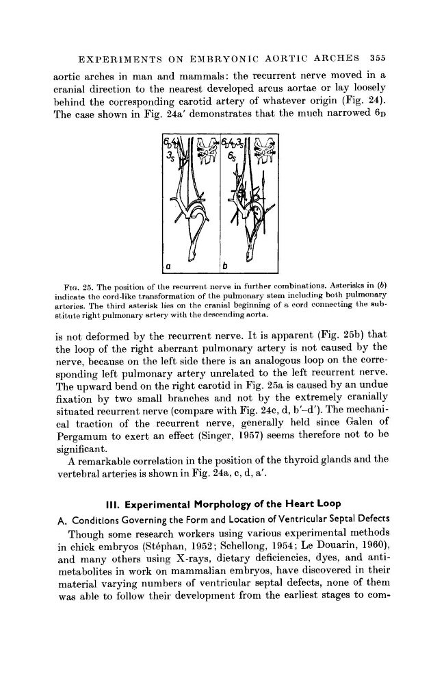
FIG. 25. The position of the recurrent nerve in further combinations. Asterisks in (6)
indicate the cord-like transformation of the pulmonary stem including both pulmonary
arteries. The third asterisk lies on the cranial beginning of a cord connecting the substitute right pulmonary artery with the descending aorta.
is not deformed by the recurrent nerve. It is apparent (Fig. 25b) that
the loop of the right aberrant pulmonary artery is not caused by the
nerve, because on the left side there is an analogous loop on the corresponding left pulmonary artery unrelated to the left recurrent nerve.
The upward bend on the right carotid in Fig. 25a is caused by an undue
fixation by two small branches and not by the extremely cranially
situated recurrent nerve (compare with Fig. 24c, d, b'-d')· The mechanical traction of the recurrent nerve, generally held since Galen of
Pergamum to exert an effect (Singer, 1957) seems therefore not to be
significant.
A remarkable correlation in the position of the thyroid glands and the
vertebral arteries is shown in Fig. 24a, c, d, a'.
III. Experimental Morphology of the Heart Loop
A. Conditions Governing the Form and Location of Ventricular Septal Defects
Though some research workers using various experimental methods
in chick embryos (Stephan, 1952; Schellong, 1954; Le Douarin, 1960),
and many others using X-rays, dietary deficiencies, dyes, and antimetabolites in work on mammalian embryos, have discovered in their
material varying numbers of ventricular septal defects, none of them
was able to follow their development from the earliest stages to com-
aortic arches in man and mammals: the recurrent nerve moved in a
cranial direction to the nearest developed arcus aortae or lay loosely
behind the corresponding carotid artery of whatever origin (Fig. 24).
The case shown in Fig. 24a' demonstrates that the much narrowed 6 D
FIG. 25. The position of the recurrent nerve in further combinations. Asterisks in (6)
indicate the cord-like transformation of the pulmonary stem including both pulmonary
arteries. The third asterisk lies on the cranial beginning of a cord connecting the substitute right pulmonary artery with the descending aorta.
is not deformed by the recurrent nerve. It is apparent (Fig. 25b) that
the loop of the right aberrant pulmonary artery is not caused by the
nerve, because on the left side there is an analogous loop on the corresponding left pulmonary artery unrelated to the left recurrent nerve.
The upward bend on the right carotid in Fig. 25a is caused by an undue
fixation by two small branches and not by the extremely cranially
situated recurrent nerve (compare with Fig. 24c, d, b'-d')· The mechanical traction of the recurrent nerve, generally held since Galen of
Pergamum to exert an effect (Singer, 1957) seems therefore not to be
significant.
A remarkable correlation in the position of the thyroid glands and the
vertebral arteries is shown in Fig. 24a, c, d, a'.
III. Experimental Morphology of the Heart Loop
A. Conditions Governing the Form and Location of Ventricular Septal Defects
Though some research workers using various experimental methods
in chick embryos (Stephan, 1952; Schellong, 1954; Le Douarin, 1960),
and many others using X-rays, dietary deficiencies, dyes, and antimetabolites in work on mammalian embryos, have discovered in their
material varying numbers of ventricular septal defects, none of them
was able to follow their development from the earliest stages to com-