

E X P E R I M E N T S ON EMBRYONIC AORTIC ARCHES 345
vessels (1919, 1921, 1923) this series might represent an increasing
persistence of the conus of the right ventricular aorta as found in
reptiles. Not trusting this seemingly plausible explanation, the problem
was approached by a new method (Rychter and Lemez, 1961b).
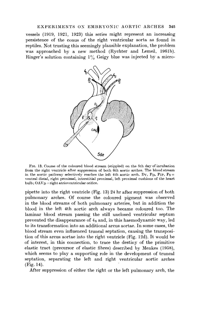
Ringer's solution containing 1% Geigy blue was injected by a microFIG. 13. Course of the coloured blood stream (stippled) on the 5th day of incubation
from the right ventricle after suppression of both 6th aortic arches. The blood stream
in the aortic pathway selectively reaches the left 4th aortic arch. Dv, ?D» PlP, Ps =
ventral distal, right proximal, interstitial proximal, left proximal cushions of the heart
bulb; O A V D = right atrioventricular orifice.
pipette into the right ventricle (Fig. 13) 24 hr after suppression of both
pulmonary arches. Of course the coloured pigment was observed
in the blood streams of both pulmonary arteries, but in addition the
blood in the left 4th aortic arch always became coloured too. The
laminar blood stream passing the still unclosed ventricular septum
prevented the disappearance of 4 S and, in this haemodynamic way, led
to its transformation into an additional arcus aortae. In some cases, the
blood stream even influenced truncal septation, causing the transposition of this arcus aortae into the right ventricle (Fig. 12d). It would be
of interest, in this connection, to trace the destiny of the primitive
elastic tract (precursor of elastic fibres) described by Menkes (1958),
which seems to play a supporting role in the development of truncal
septation, separating the left and right ventricular aortic arches
(Fig. 14).
After suppression of either the right or the left pulmonary arch, the
vessels (1919, 1921, 1923) this series might represent an increasing
persistence of the conus of the right ventricular aorta as found in
reptiles. Not trusting this seemingly plausible explanation, the problem
was approached by a new method (Rychter and Lemez, 1961b).
Ringer's solution containing 1% Geigy blue was injected by a microFIG. 13. Course of the coloured blood stream (stippled) on the 5th day of incubation
from the right ventricle after suppression of both 6th aortic arches. The blood stream
in the aortic pathway selectively reaches the left 4th aortic arch. Dv, ?D» PlP, Ps =
ventral distal, right proximal, interstitial proximal, left proximal cushions of the heart
bulb; O A V D = right atrioventricular orifice.
pipette into the right ventricle (Fig. 13) 24 hr after suppression of both
pulmonary arches. Of course the coloured pigment was observed
in the blood streams of both pulmonary arteries, but in addition the
blood in the left 4th aortic arch always became coloured too. The
laminar blood stream passing the still unclosed ventricular septum
prevented the disappearance of 4 S and, in this haemodynamic way, led
to its transformation into an additional arcus aortae. In some cases, the
blood stream even influenced truncal septation, causing the transposition of this arcus aortae into the right ventricle (Fig. 12d). It would be
of interest, in this connection, to trace the destiny of the primitive
elastic tract (precursor of elastic fibres) described by Menkes (1958),
which seems to play a supporting role in the development of truncal
septation, separating the left and right ventricular aortic arches
(Fig. 14).
After suppression of either the right or the left pulmonary arch, the