

338
Z. RYCHTER
Suppression of the left 3rd aortic arch (3s) differs from suppression of
the right in the persistence of the ventral portion of 4 S (Rychter, 1957)
as the beginning of the left carotid artery (Fig. 3).
Suppression of the right or left 6th aortic arch (6 D or 6 S ) led to the
elimination of the corresponding ductus Botalli (Fig. 3) leaving only a
slender vessel as the pulmonary artery. In some cases a short thin cord,
a persistent remnant of 4 S was present. The right 6th aortic arch was
suppressed by microelectrocoagulation (Mannhoff and Johnson, 1951)
in 2 cases out of 26 experiments and by means of X-rays (Le Douarin
and Le Douarin, 1959) in 3 out of 9 embryos, in which the 6th branchial
arch was affected. Rychter (1957) used 10 embryos for each type.
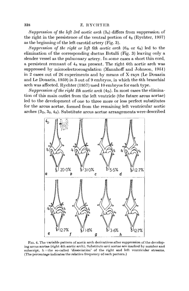
Suppression of the right 4th aortic arch (4D). In most cases the elimination of this main outlet from the left ventricle (the future arcus aortae)
led to the development of one to three more or less perfect substitutes
for the arcus aortae, formed from the remaining left ventricular aortic
arches (3 D , 3 S , 4 S ). Substitute arcus aortae arrangements were described
FIG. 4. The variable pattern of aortic arch derivatives after suppression of the developing arcus aortae (right 4th aortic arch). Substitute arci aortae are marked by number and
subscript. h = t h e so-called 'dissociation' of the right and left ventricular streams.
(The percentage indicates the relative frequency of each pattern.)
Z. RYCHTER
Suppression of the left 3rd aortic arch (3s) differs from suppression of
the right in the persistence of the ventral portion of 4 S (Rychter, 1957)
as the beginning of the left carotid artery (Fig. 3).
Suppression of the right or left 6th aortic arch (6 D or 6 S ) led to the
elimination of the corresponding ductus Botalli (Fig. 3) leaving only a
slender vessel as the pulmonary artery. In some cases a short thin cord,
a persistent remnant of 4 S was present. The right 6th aortic arch was
suppressed by microelectrocoagulation (Mannhoff and Johnson, 1951)
in 2 cases out of 26 experiments and by means of X-rays (Le Douarin
and Le Douarin, 1959) in 3 out of 9 embryos, in which the 6th branchial
arch was affected. Rychter (1957) used 10 embryos for each type.
Suppression of the right 4th aortic arch (4D). In most cases the elimination of this main outlet from the left ventricle (the future arcus aortae)
led to the development of one to three more or less perfect substitutes
for the arcus aortae, formed from the remaining left ventricular aortic
arches (3 D , 3 S , 4 S ). Substitute arcus aortae arrangements were described
FIG. 4. The variable pattern of aortic arch derivatives after suppression of the developing arcus aortae (right 4th aortic arch). Substitute arci aortae are marked by number and
subscript. h = t h e so-called 'dissociation' of the right and left ventricular streams.
(The percentage indicates the relative frequency of each pattern.)