

E X P E R I M E N T S ON EMBRYONIC AORTIC ARCHES 337
arches (3D, 3S, 4 D , 4S, 6 D , 6S—the subscript indicates the side, D =
dextral, S = sinistral). By means of clips (Fig. 2) from one to five of them
may be suppressed simultaneously in a single embryo. The 62 possible
combinations are given in Table 1.
A. Suppression of One Aortic Arch
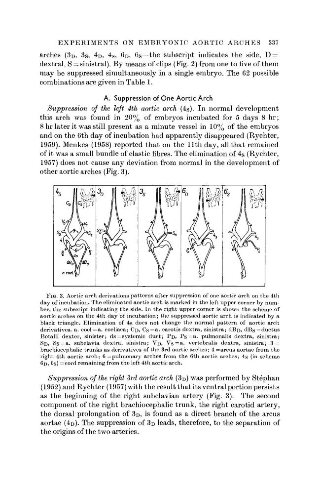
Suppression of the left 4th aortic arch (4s). In normal development
this arch was found in 20% of embryos incubated for 5 days 8 hr;
8 hr later it was still present as a minute vessel in 10% of the embryos
and on the 6th day of incubation had apparently disappeared (Rychter,
1959). Menkes (1958) reported that on the 11th day, all that remained
of it was a small bundle of elastic fibres. The elimination of 4 S (Rychter,
1957) does not cause any deviation from normal in the development of
other aortic arches (Fig. 3).
FIG. 3. Aortic arch derivations patterns after suppression of one aortic arch on the 4th
day of incubation. The eliminated aortic arch is marked in the left upper corner by number, the subscript indicating the side. In the right upper corner is shown the scheme of
aortic arches on the 4th day of incubation; the suppressed aortic arch is indicated by a
black triangle. Elimination of 4s does not change the normal pattern of aortic arch
derivatives, a. coel=a. coeliaca; CD, C s = a . carotis dextra, sinistra; dBi>, d B s = d u c t u s
Botalli dexter, sinister; d s = systemic duct; P D , P s = a . pulmonalis dextra, sinistra;
$D> S s = a . subclavia dextra, sinistra; V D , V s = a . vertebralis dextra, sinistra; 3 =
brachiocephalic trunks as derivatives of the 3rd aortic arches; 4 = arcus aortae from the
right 4th aortic arch; 6 = pulmonary arches from the 6th aortic arches; 4s (in scheme
6D> 6S) =cord remaining from the left 4th aortic arch.
Suppression of the right 3rd aortic arch (3D) was performed by Stephan
(1952) and Rychter (1957) with the result that its ventral portion persist s
as the beginning of the right subclavian artery (Fig. 3). The second
component of the right brachiocephalic trunk, the right carotid artery,
the dorsal prolongation of 3D, is found as a direct branch of the arcus
aortae (4D). The suppression of 3D leads, therefore, to the separation of
the origins of the two arteries.
arches (3D, 3S, 4 D , 4S, 6 D , 6S—the subscript indicates the side, D =
dextral, S = sinistral). By means of clips (Fig. 2) from one to five of them
may be suppressed simultaneously in a single embryo. The 62 possible
combinations are given in Table 1.
A. Suppression of One Aortic Arch
Suppression of the left 4th aortic arch (4s). In normal development
this arch was found in 20% of embryos incubated for 5 days 8 hr;
8 hr later it was still present as a minute vessel in 10% of the embryos
and on the 6th day of incubation had apparently disappeared (Rychter,
1959). Menkes (1958) reported that on the 11th day, all that remained
of it was a small bundle of elastic fibres. The elimination of 4 S (Rychter,
1957) does not cause any deviation from normal in the development of
other aortic arches (Fig. 3).
FIG. 3. Aortic arch derivations patterns after suppression of one aortic arch on the 4th
day of incubation. The eliminated aortic arch is marked in the left upper corner by number, the subscript indicating the side. In the right upper corner is shown the scheme of
aortic arches on the 4th day of incubation; the suppressed aortic arch is indicated by a
black triangle. Elimination of 4s does not change the normal pattern of aortic arch
derivatives, a. coel=a. coeliaca; CD, C s = a . carotis dextra, sinistra; dBi>, d B s = d u c t u s
Botalli dexter, sinister; d s = systemic duct; P D , P s = a . pulmonalis dextra, sinistra;
$D> S s = a . subclavia dextra, sinistra; V D , V s = a . vertebralis dextra, sinistra; 3 =
brachiocephalic trunks as derivatives of the 3rd aortic arches; 4 = arcus aortae from the
right 4th aortic arch; 6 = pulmonary arches from the 6th aortic arches; 4s (in scheme
6D> 6S) =cord remaining from the left 4th aortic arch.
Suppression of the right 3rd aortic arch (3D) was performed by Stephan
(1952) and Rychter (1957) with the result that its ventral portion persist s
as the beginning of the right subclavian artery (Fig. 3). The second
component of the right brachiocephalic trunk, the right carotid artery,
the dorsal prolongation of 3D, is found as a direct branch of the arcus
aortae (4D). The suppression of 3D leads, therefore, to the separation of
the origins of the two arteries.