

334
Z. RYCHTER
ong, 1954), X-rays (e.g., Wilson and Karr, 1951). The malformations
obtained were fully comparable with those occurring in man.
With these experimental arrangements it was impossible to follow
either the morphological development (because of the irregular
occurrence and variable form) or the developmental mechanism that
produces the malformation. Nowadays, the developmental mechanism
is, in our opinion, the most important problem to be investigated for
further progress to be made in experimental teratology. The study of
developmental mechanisms requires a series of embryos at successive
developmental stages having the same malformation. Such material can
be provided only by experimental intervention at a particular site in
the embryo. Only those papers, therefore, in which such local intervention was attempted, will be reviewed here.
1. Experiments on Aortic Arches
a. Birds
By cauterization of the lateral side of chick embryos, in order to
obtain reversed body flexion, Bremer (1928) tried to change the pattern
of aortic arches. Results on three successfully cauterized embryos were
uninterpretable. Hinrichs (1931), using localized U.V. irradiation,
succeeded in suppressing all the right side aortic arches. Unfortunately
she did not continue her experiments. MannhofF and Johnson (1951)
applied localized microcoagulation to the right 6th aortic arch, obtaining in most cases only stenosis of this vessel. Stephan (1952) mentions
the use of X-rays in an attempt to suppress aortic arch development in
chick embryos. Inconsistent results (which are also apparent in the
paper of Le Douarin and Le Douarin, 1959) persuaded him to work
solely on mechanical suppression of the aortic arches by means of a
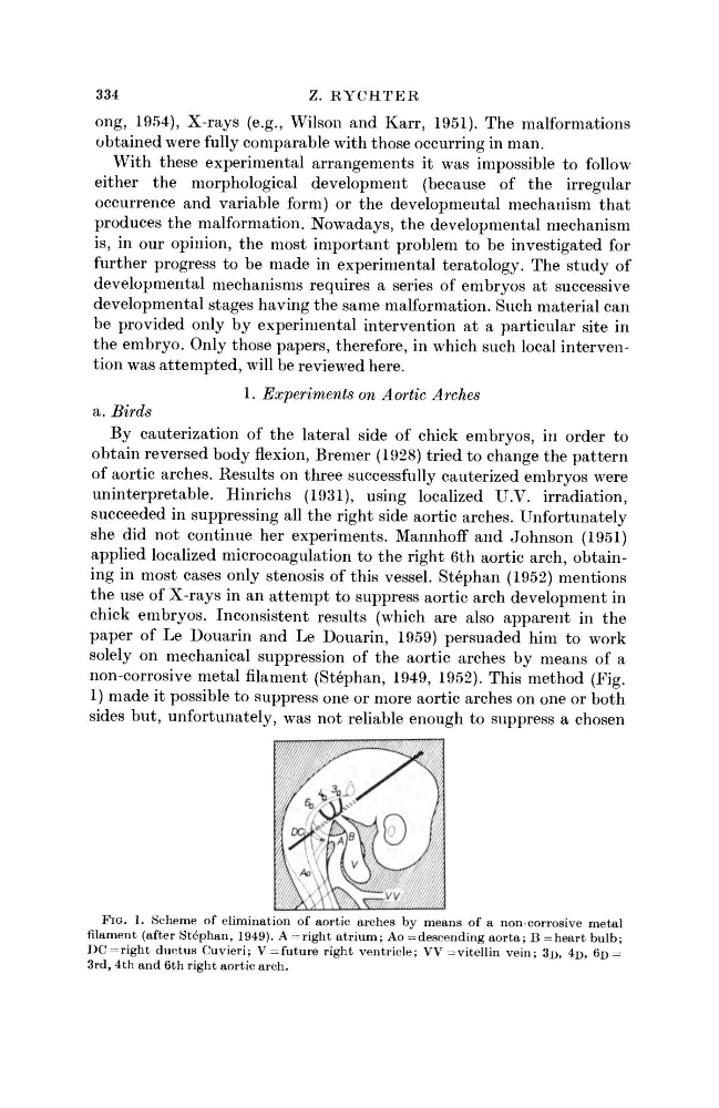
non-corrosive metal filament (Stephan, 1949, 1952). This method (Fig.
1) made it possible to suppress one or more aortic arches on one or both
sides but, unfortunately, was not reliable enough to suppress a chosen
FIG. 1. Scheme of elimination of aortic arches by means of a non-corrosive metal
filament (after Stephan, 1949). A = right atrium; Ao = descending aorta; B = heart bulb;
DC = right ductus Cuvieri; V = future right ventricle; W = v i t e l l i n vein; 3D, 4 D , 6D =
3rd, 4th and 6th right aortic arch.
Z. RYCHTER
ong, 1954), X-rays (e.g., Wilson and Karr, 1951). The malformations
obtained were fully comparable with those occurring in man.
With these experimental arrangements it was impossible to follow
either the morphological development (because of the irregular
occurrence and variable form) or the developmental mechanism that
produces the malformation. Nowadays, the developmental mechanism
is, in our opinion, the most important problem to be investigated for
further progress to be made in experimental teratology. The study of
developmental mechanisms requires a series of embryos at successive
developmental stages having the same malformation. Such material can
be provided only by experimental intervention at a particular site in
the embryo. Only those papers, therefore, in which such local intervention was attempted, will be reviewed here.
1. Experiments on Aortic Arches
a. Birds
By cauterization of the lateral side of chick embryos, in order to
obtain reversed body flexion, Bremer (1928) tried to change the pattern
of aortic arches. Results on three successfully cauterized embryos were
uninterpretable. Hinrichs (1931), using localized U.V. irradiation,
succeeded in suppressing all the right side aortic arches. Unfortunately
she did not continue her experiments. MannhofF and Johnson (1951)
applied localized microcoagulation to the right 6th aortic arch, obtaining in most cases only stenosis of this vessel. Stephan (1952) mentions
the use of X-rays in an attempt to suppress aortic arch development in
chick embryos. Inconsistent results (which are also apparent in the
paper of Le Douarin and Le Douarin, 1959) persuaded him to work
solely on mechanical suppression of the aortic arches by means of a
non-corrosive metal filament (Stephan, 1949, 1952). This method (Fig.
1) made it possible to suppress one or more aortic arches on one or both
sides but, unfortunately, was not reliable enough to suppress a chosen
FIG. 1. Scheme of elimination of aortic arches by means of a non-corrosive metal
filament (after Stephan, 1949). A = right atrium; Ao = descending aorta; B = heart bulb;
DC = right ductus Cuvieri; V = future right ventricle; W = v i t e l l i n vein; 3D, 4 D , 6D =
3rd, 4th and 6th right aortic arch.