

308
TH. LENDER
Dugesia lugubris and by Stephan-Dubois (1961) on Dendrocoelum
lacteum. In the former, the neoblasts are found throughout the body of
the planarian, occurring with greater frequency along the nerve trunks.
This is also true of Planaria vitta (Pedersen, 1959b). The neoblasts
decrease in number from the head towards the tail. In Dugesia lugubris
they are distributed according to a decreasing gradient from front to
back with two maxima, one in the region of the brain, the other in the
prepharyngeal region (Fig. 2). During periods of starvation neoblasts
can persist in the planarians for two months. In Dendrocoelum lacteum,
the neoblasts are sparser and more uniformly distributed (StephanDubois, 1961).
B. Neoblasts of the Regeneration Blastema
Neoblasts are found in the regeneration blastemas. Pedersen (1959b)
has photographed them in vivo through the phase contrast microscope.
Methyl green-pyronine staining shows that these cells are identical
with the neoblasts in the body of the planarians. In all instances, even in
Planaria after two months of starvation, blastemas are built up by
accumulation of neoblasts.
What is the origin of the neoblasts of the blastema?
They may be considered to arise either from the dedifferentiation of
the tissues of the wound or from the migration of the neoblasts of the
planarian body toward the wound. The investigations of Wolff and
Dubois (1947a, b, c) and of Dubois (1949) support the latter hypothesis.
The formation of a regeneration blastema is the result of the division
and migration of the neoblasts. This process is started and directed by a
stimulus emitted by the wound.
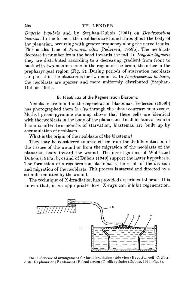
The technique of X-irradiation has provided experimental proof. It is
known that, in an appropriate dose, X-rays can inhibit regeneration.
FIG. 3. Scheme of arrangement for local irradiation (side view) B : cotton coil; C: Petri
dish; D : planarian; F : filament; P : lead screen; T: silk cylinder (Dubois, 1949, Fig. 3).
TH. LENDER
Dugesia lugubris and by Stephan-Dubois (1961) on Dendrocoelum
lacteum. In the former, the neoblasts are found throughout the body of
the planarian, occurring with greater frequency along the nerve trunks.
This is also true of Planaria vitta (Pedersen, 1959b). The neoblasts
decrease in number from the head towards the tail. In Dugesia lugubris
they are distributed according to a decreasing gradient from front to
back with two maxima, one in the region of the brain, the other in the
prepharyngeal region (Fig. 2). During periods of starvation neoblasts
can persist in the planarians for two months. In Dendrocoelum lacteum,
the neoblasts are sparser and more uniformly distributed (StephanDubois, 1961).
B. Neoblasts of the Regeneration Blastema
Neoblasts are found in the regeneration blastemas. Pedersen (1959b)
has photographed them in vivo through the phase contrast microscope.
Methyl green-pyronine staining shows that these cells are identical
with the neoblasts in the body of the planarians. In all instances, even in
Planaria after two months of starvation, blastemas are built up by
accumulation of neoblasts.
What is the origin of the neoblasts of the blastema?
They may be considered to arise either from the dedifferentiation of
the tissues of the wound or from the migration of the neoblasts of the
planarian body toward the wound. The investigations of Wolff and
Dubois (1947a, b, c) and of Dubois (1949) support the latter hypothesis.
The formation of a regeneration blastema is the result of the division
and migration of the neoblasts. This process is started and directed by a
stimulus emitted by the wound.
The technique of X-irradiation has provided experimental proof. It is
known that, in an appropriate dose, X-rays can inhibit regeneration.
FIG. 3. Scheme of arrangement for local irradiation (side view) B : cotton coil; C: Petri
dish; D : planarian; F : filament; P : lead screen; T: silk cylinder (Dubois, 1949, Fig. 3).