

22
V A N C E T A R T A R
characteristic of that of the nucleus, as in Acetabularia, we cannot say
because the feeding organelles of stentors appear so much alike. It is
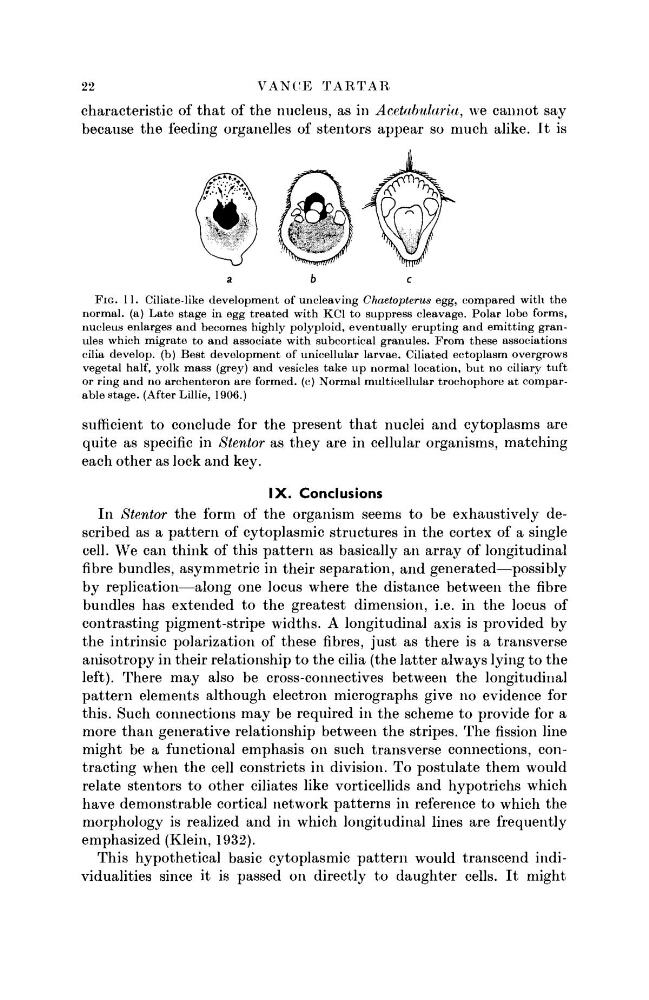
FIG. 11. Ciliate-like development of uncleaving Chaetopterus egg, compared with the
normal, (a) Late stage in egg treated with KC1 to suppress cleavage. Polar lobe forms,
nucleus enlarges and becomes highly polyploid, eventually erupting and emitting granules which migrate to and associate with subcortical granules. From these associations
cilia develop, (b) Best development of unicellular larvae. Ciliated ectoplasm overgrows
vegetal half, yolk mass (grey) and vesicles take up normal location, but no ciliary tuft
or ring and no archenteron are formed, (c) Normal multicellular trochophore at comparable stage. (After Lillie, 1906.)
sufficient to conclude for the present that nuclei and cytoplasms are
quite as specific in Stentor as they are in cellular organisms, matching
each other as lock and key.
I X . Conclusions
In Stentor the form of the organism seems to be exhaustively described as a pattern of cytoplasmic structures in the cortex of a single
cell. We can think of this pattern as basically an array of longitudinal
fibre bundles, asymmetric in their separation, and generated—possibly
by replication—along one locus where the distance between the fibre
bundles has extended to the greatest dimension, i.e. in the locus of
contrasting pigment-stripe widths. A longitudinal axis is provided by
the intrinsic polarization of these fibres, just as there is a transverse
anisotropy in their relationship to the cilia (the latter always lying to the
left). There may also be cross-connectives between the longitudinal
pattern elements although electron micrographs give no evidence for
this. Such connections may be required in the scheme to provide for a
more than generative relationship between the stripes. The fission line
might be a functional emphasis on such transverse connections, contracting when the cell constricts in division. To postulate them would
relate stentors to other ciliates like vorticellids and hypotrichs which
have demonstrable cortical network patterns in reference to which the
morphology is realized and in which longitudinal lines are frequently
emphasized (Klein, 1932).
This hypothetical basic cytoplasmic pattern would transcend individualities since it is passed on directly to daughter cells. It might
V A N C E T A R T A R
characteristic of that of the nucleus, as in Acetabularia, we cannot say
because the feeding organelles of stentors appear so much alike. It is
FIG. 11. Ciliate-like development of uncleaving Chaetopterus egg, compared with the
normal, (a) Late stage in egg treated with KC1 to suppress cleavage. Polar lobe forms,
nucleus enlarges and becomes highly polyploid, eventually erupting and emitting granules which migrate to and associate with subcortical granules. From these associations
cilia develop, (b) Best development of unicellular larvae. Ciliated ectoplasm overgrows
vegetal half, yolk mass (grey) and vesicles take up normal location, but no ciliary tuft
or ring and no archenteron are formed, (c) Normal multicellular trochophore at comparable stage. (After Lillie, 1906.)
sufficient to conclude for the present that nuclei and cytoplasms are
quite as specific in Stentor as they are in cellular organisms, matching
each other as lock and key.
I X . Conclusions
In Stentor the form of the organism seems to be exhaustively described as a pattern of cytoplasmic structures in the cortex of a single
cell. We can think of this pattern as basically an array of longitudinal
fibre bundles, asymmetric in their separation, and generated—possibly
by replication—along one locus where the distance between the fibre
bundles has extended to the greatest dimension, i.e. in the locus of
contrasting pigment-stripe widths. A longitudinal axis is provided by
the intrinsic polarization of these fibres, just as there is a transverse
anisotropy in their relationship to the cilia (the latter always lying to the
left). There may also be cross-connectives between the longitudinal
pattern elements although electron micrographs give no evidence for
this. Such connections may be required in the scheme to provide for a
more than generative relationship between the stripes. The fission line
might be a functional emphasis on such transverse connections, contracting when the cell constricts in division. To postulate them would
relate stentors to other ciliates like vorticellids and hypotrichs which
have demonstrable cortical network patterns in reference to which the
morphology is realized and in which longitudinal lines are frequently
emphasized (Klein, 1932).
This hypothetical basic cytoplasmic pattern would transcend individualities since it is passed on directly to daughter cells. It might