

240
S I L V I O R A N Z I
to protein denaturation. As already mentioned, urea was effective in
inducing animalization in the sea urchin embryo (Pedrazzi, 1957) and
in inducing formation of notochord from the ventral half of the frog
gastrula (Leone, 1952). We may therefore conclude that protein denaturation is one of the steps in animalization.
Ranzi (1942) pointed out that all the animalizing substances are
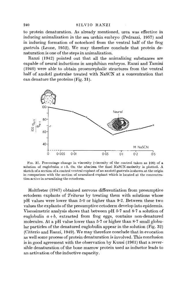
capable of neural inductions in amphibian embryos. Ranzi and Tamini
(1940) were able to obtain prosencephalic structures from the ventral
half of axolotl gastrulae treated with NaSCN at a concentration that
can denature the proteins (Fig. 31).
Neural
FIG. 31. Percentage change in viscosity (viscosity of the control taken as 100) of a
solution of euglobulin a+b. On the abscissa the final NaSCN-molarity is plotted. A
sketch of a section of a control ventral explant of an axolotl gastrula is shown at the origin
in comparison with the section of neuralized explant which is located at the concentration active in neuralizing the ectoderm.
Holtfreter (1947) obtained nervous differentiation from presumptive
ectoderm explants of Triturus by treating them with solutions whose
pH values were lower than 5-0 or higher than 9-2. Between these two
values the explants of the presumptive ectoderm develop into epidermis.
Viscosimetric analysis shows that between pH 5-7 and 8-7 a solution of
euglobulin a + 6, extracted from frog eggs, contains non-denatured
molecules. At a pH value lower than 5-7 or higher than 8-7 small globular particles of the denatured euglobulin appear in the solution (Fig. 32)
(Citterio and Ranzi, 1949). We may therefore conclude that in evocation
as well some process of protein denaturation is involved. This conclusion
is in good agreement with the observation by Kuusi (1961) that a reversible denaturation of the bone marrow protein used as inductor leads to
an activation of the inductive capacity.
S I L V I O R A N Z I
to protein denaturation. As already mentioned, urea was effective in
inducing animalization in the sea urchin embryo (Pedrazzi, 1957) and
in inducing formation of notochord from the ventral half of the frog
gastrula (Leone, 1952). We may therefore conclude that protein denaturation is one of the steps in animalization.
Ranzi (1942) pointed out that all the animalizing substances are
capable of neural inductions in amphibian embryos. Ranzi and Tamini
(1940) were able to obtain prosencephalic structures from the ventral
half of axolotl gastrulae treated with NaSCN at a concentration that
can denature the proteins (Fig. 31).
Neural
FIG. 31. Percentage change in viscosity (viscosity of the control taken as 100) of a
solution of euglobulin a+b. On the abscissa the final NaSCN-molarity is plotted. A
sketch of a section of a control ventral explant of an axolotl gastrula is shown at the origin
in comparison with the section of neuralized explant which is located at the concentration active in neuralizing the ectoderm.
Holtfreter (1947) obtained nervous differentiation from presumptive
ectoderm explants of Triturus by treating them with solutions whose
pH values were lower than 5-0 or higher than 9-2. Between these two
values the explants of the presumptive ectoderm develop into epidermis.
Viscosimetric analysis shows that between pH 5-7 and 8-7 a solution of
euglobulin a + 6, extracted from frog eggs, contains non-denatured
molecules. At a pH value lower than 5-7 or higher than 8-7 small globular particles of the denatured euglobulin appear in the solution (Fig. 32)
(Citterio and Ranzi, 1949). We may therefore conclude that in evocation
as well some process of protein denaturation is involved. This conclusion
is in good agreement with the observation by Kuusi (1961) that a reversible denaturation of the bone marrow protein used as inductor leads to
an activation of the inductive capacity.