

P R O T E I N S I N D E V E L O P M E N T
239
globular particles and induces a reduction in the anisotropy of the
fibrillar particles. This phenomenon was directly observed with the
electron microscope on actomyosin treated with SCN~ and I
- (Ranzi,
0 0-005 0-01
005
0-1
0-2
θ'·5 M SCN o 7 ü C l
6
CH
05
ί I0~
3 M IBA 3
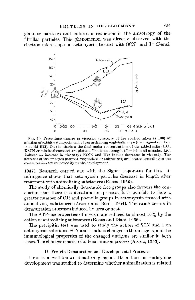
FIG. 30. Percentage change in viscosity (viscosity of the control taken as 100) of
solution of rabbit actomyosin and of sea urchin egg euglobulin a + b (the original solution
is in IM KC1). On the abscissa the final molar concentrations of the added salts (LiCl,
KSCN or o-iodosobenzoate) are plotted. The ionic strength (I) =1-0 in all samples. LiCl
induces an increase in viscosity; KSCN and IBA induce decreases in viscosity. The
sketches of the embryos (normal, vegetalized or animalized) are located according to the
concentration active in modifying the development.
1947). Research carried out with the Signer apparatus for flow birefringence shows that actomyosin particles decrease in length after
treatment with animalizing substances (Rocca, 1956).
The study of chemically detectable free groups also favours the conclusion that there is a denaturation process. It is possible to show a
greater number of OH and phenolic groups in actomyosin treated with
animalizing substances (Arosio and Bossi, 1954). The same occurs in
denaturation processes induced by urea or heat.
The ATP-ase properties of myosin are reduced to almost 10% by the
action of animalizing substances (Rocca and Diani, 1956).
The precipitin test was used to study the action of SCN and I on
actomyosin solutions. SCN and I induce changes in the antigens, and the
immunological properties of the changed antigens are similar in both
cases. The changes consist of a denaturation process (Arosio, 1953).
D. Protein Denaturation and Developmental Processes
Urea is a well-known denaturing agent. Its action on embryonic
development was studied to determine whether animalization is related
239
globular particles and induces a reduction in the anisotropy of the
fibrillar particles. This phenomenon was directly observed with the
electron microscope on actomyosin treated with SCN~ and I
- (Ranzi,
0 0-005 0-01
005
0-1
0-2
θ'·5 M SCN o 7 ü C l
6
CH
05
ί I0~
3 M IBA 3
FIG. 30. Percentage change in viscosity (viscosity of the control taken as 100) of
solution of rabbit actomyosin and of sea urchin egg euglobulin a + b (the original solution
is in IM KC1). On the abscissa the final molar concentrations of the added salts (LiCl,
KSCN or o-iodosobenzoate) are plotted. The ionic strength (I) =1-0 in all samples. LiCl
induces an increase in viscosity; KSCN and IBA induce decreases in viscosity. The
sketches of the embryos (normal, vegetalized or animalized) are located according to the
concentration active in modifying the development.
1947). Research carried out with the Signer apparatus for flow birefringence shows that actomyosin particles decrease in length after
treatment with animalizing substances (Rocca, 1956).
The study of chemically detectable free groups also favours the conclusion that there is a denaturation process. It is possible to show a
greater number of OH and phenolic groups in actomyosin treated with
animalizing substances (Arosio and Bossi, 1954). The same occurs in
denaturation processes induced by urea or heat.
The ATP-ase properties of myosin are reduced to almost 10% by the
action of animalizing substances (Rocca and Diani, 1956).
The precipitin test was used to study the action of SCN and I on
actomyosin solutions. SCN and I induce changes in the antigens, and the
immunological properties of the changed antigens are similar in both
cases. The changes consist of a denaturation process (Arosio, 1953).
D. Protein Denaturation and Developmental Processes
Urea is a well-known denaturing agent. Its action on embryonic
development was studied to determine whether animalization is related