

MORPHOGENESIS IN STENTOR
17
tive morphogenetic tendencies are eventually manifested as both
doublets and triplets transform to the single normal form, accomplished
by the obliteration of supernumerary mouthparts and primordium sites.
V I I . Reconstitution of Minced Stentor
Stentors can be cut into repeatedly with a glass needle until the ectoplasmic structure consists of forty or more stripe patches too small for
further cutting and lying in random arrangement. When the head and
tail are first excised, the Stentor is left in the maximum state of anisotropy obtainable. The original cell axis is completely obliterated and the
normal pattern of the striping is entirely disrupted. Yet minced stentors
regenerate and can reconstitute the normal form within a day. This
phenomenon is in many ways analogous to the reconstitution of sponges
and hydroids from dissociated cells, as well as tissue formation in
embryonic cell aggregates.
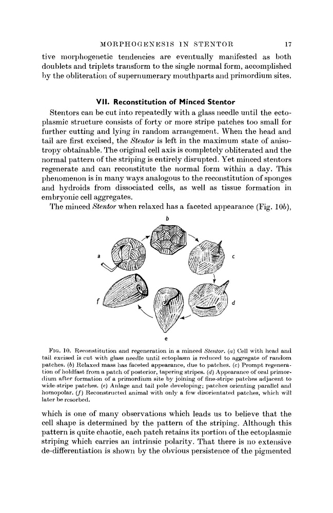
The minced Stentor when relaxed has a faceted appearance (Fig. 106),
FIG. 10. Reconstitution and regeneration in a minced Stentor. (a) Cell with head and
tail excised is cut with glass needle until ectoplasm is reduced to aggregate of random
patches, (b) Relaxed mass has faceted appearance, due to patches, (c) Prompt regeneration of holdfast from a patch of posterior, tapering stripes, (d) Appearance of oral primordium after formation of a primordium site by joining of fine-stripe patches adjacent to
wide-stripe patches, (e) Anlage and tail pole developing; patches orienting parallel and
homopolar. (/) Reconstructed animal with only a few disorientated patches, which will
later be resorbed.
which is one of many observations which leads us to believe that the
cell shape is determined by the pattern of the striping. Although this
pattern is quite chaotic, each patch retains its portion of the ectoplasmic
striping which carries an intrinsic polarity. That there is no extensive
de-differentiation is shown by the obvious persistence of the pigmented
17
tive morphogenetic tendencies are eventually manifested as both
doublets and triplets transform to the single normal form, accomplished
by the obliteration of supernumerary mouthparts and primordium sites.
V I I . Reconstitution of Minced Stentor
Stentors can be cut into repeatedly with a glass needle until the ectoplasmic structure consists of forty or more stripe patches too small for
further cutting and lying in random arrangement. When the head and
tail are first excised, the Stentor is left in the maximum state of anisotropy obtainable. The original cell axis is completely obliterated and the
normal pattern of the striping is entirely disrupted. Yet minced stentors
regenerate and can reconstitute the normal form within a day. This
phenomenon is in many ways analogous to the reconstitution of sponges
and hydroids from dissociated cells, as well as tissue formation in
embryonic cell aggregates.
The minced Stentor when relaxed has a faceted appearance (Fig. 106),
FIG. 10. Reconstitution and regeneration in a minced Stentor. (a) Cell with head and
tail excised is cut with glass needle until ectoplasm is reduced to aggregate of random
patches, (b) Relaxed mass has faceted appearance, due to patches, (c) Prompt regeneration of holdfast from a patch of posterior, tapering stripes, (d) Appearance of oral primordium after formation of a primordium site by joining of fine-stripe patches adjacent to
wide-stripe patches, (e) Anlage and tail pole developing; patches orienting parallel and
homopolar. (/) Reconstructed animal with only a few disorientated patches, which will
later be resorbed.
which is one of many observations which leads us to believe that the
cell shape is determined by the pattern of the striping. Although this
pattern is quite chaotic, each patch retains its portion of the ectoplasmic
striping which carries an intrinsic polarity. That there is no extensive
de-differentiation is shown by the obvious persistence of the pigmented