

16
VANCE TARTAR
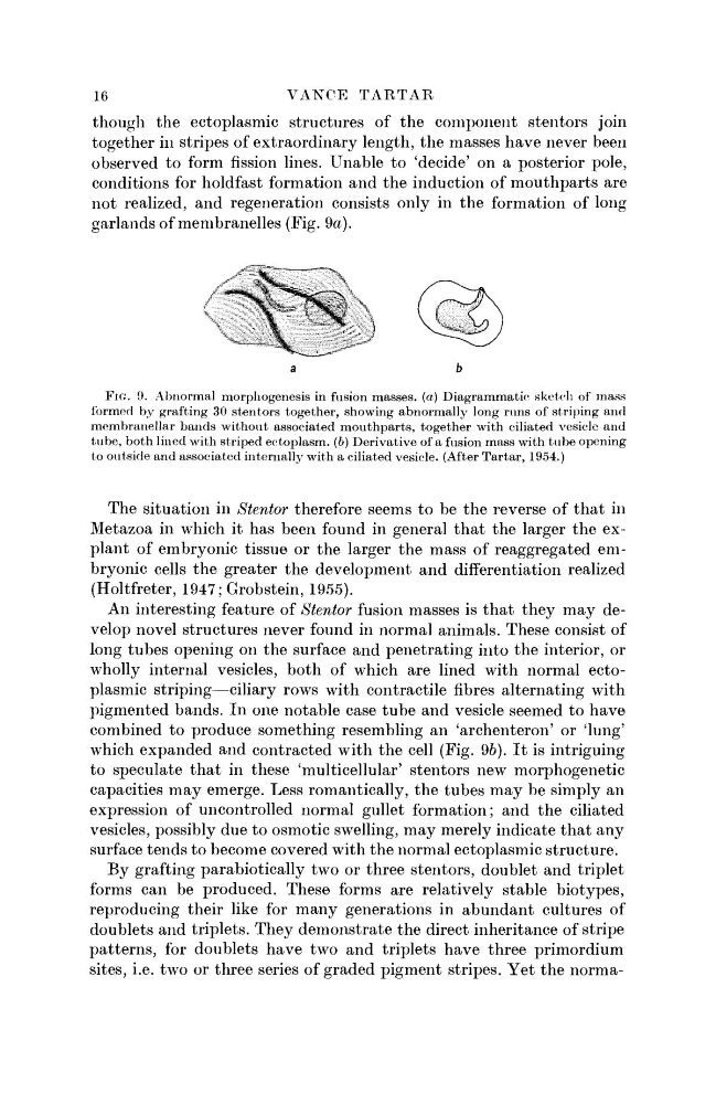
though the ectoplasmic structures of the component stentors join
together in stripes of extraordinary length, the masses have never been
observed to form fission lines. Unable to 'decide' on a posterior pole,
conditions for holdfast formation and the induction of mouthparts are
not realized, and regeneration consists only in the formation of long
garlands of membranelies (Fig. 9a).
FIG. 9. Abnormal morphogenesis in fusion masses, (a) Diagrammatic sketch of mass
formed by grafting 30 stentors together, showing abnormally long runs of striping and
membranellar bands without associated mouthparts, together with ciliated vesicle and
tube, both lined with striped ectoplasm, (b) Derivative of a fusion mass with tube opening
to outside and associated internally with a ciliated vesicle. (After Tartar, 1954.)
The situation in Stentor therefore seems to be the reverse of that in
Metazoa in which it has been found in general that the larger the explant of embryonic tissue or the larger the mass of reaggregated embryonic cells the greater the development and differentiation realized
(Holtfreter, 1947; Grobstein, 1955).
An interesting feature of Stentor fusion masses is that they may develop novel structures never found in normal animals. These consist of
long tubes opening on the surface and penetrating into the interior, or
wholly internal vesicles, both of which are lined with normal ectoplasmic striping—ciliary rows with contractile fibres alternating with
pigmented bands. In one notable case tube and vesicle seemed to have
combined to produce something resembling an 'archenteron' or 'lung'
which expanded and contracted with the cell (Fig. 96). It is intriguing
to speculate that in these 'multicellular' stentors new morphogenetic
capacities may emerge. Less romantically, the tubes may be simply an
expression of uncontrolled normal gullet formation; and the ciliated
vesicles, possibly due to osmotic swelling, may merely indicate that any
surface tends to become covered with the normal ectoplasmic structure.
By grafting parabiotically two or three stentors, doublet and triplet
forms can be produced. These forms are relatively stable biotypes,
reproducing their like for many generations in abundant cultures of
doublets and triplets. They demonstrate the direct inheritance of stripe
patterns, for doublets have two and triplets have three primordium
sites, i.e. two or three series of graded pigment stripes. Yet the norma-
VANCE TARTAR
though the ectoplasmic structures of the component stentors join
together in stripes of extraordinary length, the masses have never been
observed to form fission lines. Unable to 'decide' on a posterior pole,
conditions for holdfast formation and the induction of mouthparts are
not realized, and regeneration consists only in the formation of long
garlands of membranelies (Fig. 9a).
FIG. 9. Abnormal morphogenesis in fusion masses, (a) Diagrammatic sketch of mass
formed by grafting 30 stentors together, showing abnormally long runs of striping and
membranellar bands without associated mouthparts, together with ciliated vesicle and
tube, both lined with striped ectoplasm, (b) Derivative of a fusion mass with tube opening
to outside and associated internally with a ciliated vesicle. (After Tartar, 1954.)
The situation in Stentor therefore seems to be the reverse of that in
Metazoa in which it has been found in general that the larger the explant of embryonic tissue or the larger the mass of reaggregated embryonic cells the greater the development and differentiation realized
(Holtfreter, 1947; Grobstein, 1955).
An interesting feature of Stentor fusion masses is that they may develop novel structures never found in normal animals. These consist of
long tubes opening on the surface and penetrating into the interior, or
wholly internal vesicles, both of which are lined with normal ectoplasmic striping—ciliary rows with contractile fibres alternating with
pigmented bands. In one notable case tube and vesicle seemed to have
combined to produce something resembling an 'archenteron' or 'lung'
which expanded and contracted with the cell (Fig. 96). It is intriguing
to speculate that in these 'multicellular' stentors new morphogenetic
capacities may emerge. Less romantically, the tubes may be simply an
expression of uncontrolled normal gullet formation; and the ciliated
vesicles, possibly due to osmotic swelling, may merely indicate that any
surface tends to become covered with the normal ectoplasmic structure.
By grafting parabiotically two or three stentors, doublet and triplet
forms can be produced. These forms are relatively stable biotypes,
reproducing their like for many generations in abundant cultures of
doublets and triplets. They demonstrate the direct inheritance of stripe
patterns, for doublets have two and triplets have three primordium
sites, i.e. two or three series of graded pigment stripes. Yet the norma-