

304
Β. Μ. SHAFFER
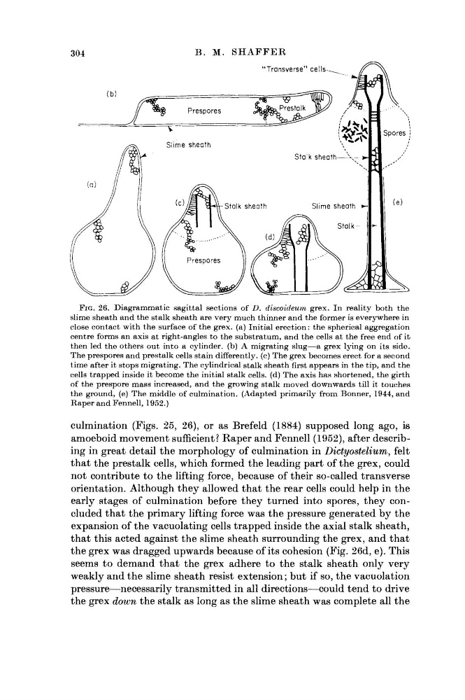
FIG. 26. Diagrammatic sagittal sections of D. discoideum grex. In reality both the
slime sheath and the stalk sheath are very much thinner and the former is everywhere in
close contact with the surface of the grex. (a) Initial erection: the spherical aggregation
centre forms an axis at right-angles to the substratum, and the cells at the free end of it
then led the others out into a cylinder, (b) A migrating slug—a grex lying on its side.
The prespores and prestalk cells stain differently, (c) The grex becomes erect for a second
time after it stops migrating. The cylindrical stalk sheath first appears in the tip, and the
cells trapped inside it become the initial stalk cells, (d) The axis has shortened, the girth
of the prespore mass increased, and the growing stalk moved downwards till it touches
the ground, (e) The middle of culmination. (Adapted primarily from Bonner, 1944, and
Raper and Fennell, 1952.)
culmination (Figs. 25, 26), or as Brefeld (1884) supposed long ago, is
amoeboid movement sufficient? Raper and Fennell (1952), after describing in great detail the morphology of culmination in Dictyostelium,
felt
that the prestalk cells, which formed the leading part of the grex, could
not contribute to the lifting force, because of their so-called transverse
orientation. Although they allowed that the rear cells could help in the
early stages of culmination before they turned into spores, they concluded that the primary lifting force was the pressure generated by the
expansion of the vacuolating cells trapped inside the axial stalk sheath,
that this acted against the slime sheath surrounding the grex, and that
the grex was dragged upwards because of its cohesion (Fig. 26d, e). This
seems to demand that the grex adhere to the stalk sheath only very
weakly and the slime sheath resist extension ; but if so, the vacuolation
pressure—necessarily transmitted in all directions—could tend to drive
the grex down the stalk as long as the slime sheath was complete all the
Β. Μ. SHAFFER
FIG. 26. Diagrammatic sagittal sections of D. discoideum grex. In reality both the
slime sheath and the stalk sheath are very much thinner and the former is everywhere in
close contact with the surface of the grex. (a) Initial erection: the spherical aggregation
centre forms an axis at right-angles to the substratum, and the cells at the free end of it
then led the others out into a cylinder, (b) A migrating slug—a grex lying on its side.
The prespores and prestalk cells stain differently, (c) The grex becomes erect for a second
time after it stops migrating. The cylindrical stalk sheath first appears in the tip, and the
cells trapped inside it become the initial stalk cells, (d) The axis has shortened, the girth
of the prespore mass increased, and the growing stalk moved downwards till it touches
the ground, (e) The middle of culmination. (Adapted primarily from Bonner, 1944, and
Raper and Fennell, 1952.)
culmination (Figs. 25, 26), or as Brefeld (1884) supposed long ago, is
amoeboid movement sufficient? Raper and Fennell (1952), after describing in great detail the morphology of culmination in Dictyostelium,
felt
that the prestalk cells, which formed the leading part of the grex, could
not contribute to the lifting force, because of their so-called transverse
orientation. Although they allowed that the rear cells could help in the
early stages of culmination before they turned into spores, they concluded that the primary lifting force was the pressure generated by the
expansion of the vacuolating cells trapped inside the axial stalk sheath,
that this acted against the slime sheath surrounding the grex, and that
the grex was dragged upwards because of its cohesion (Fig. 26d, e). This
seems to demand that the grex adhere to the stalk sheath only very
weakly and the slime sheath resist extension ; but if so, the vacuolation
pressure—necessarily transmitted in all directions—could tend to drive
the grex down the stalk as long as the slime sheath was complete all the