

EARLY DEVELOPMENT OF THE SEA URCHIN
115
beyond the stage of the triradiate spicules and in the case of aggregates
from pluteal cells no skeleton at all is formed. It is interesting to compare
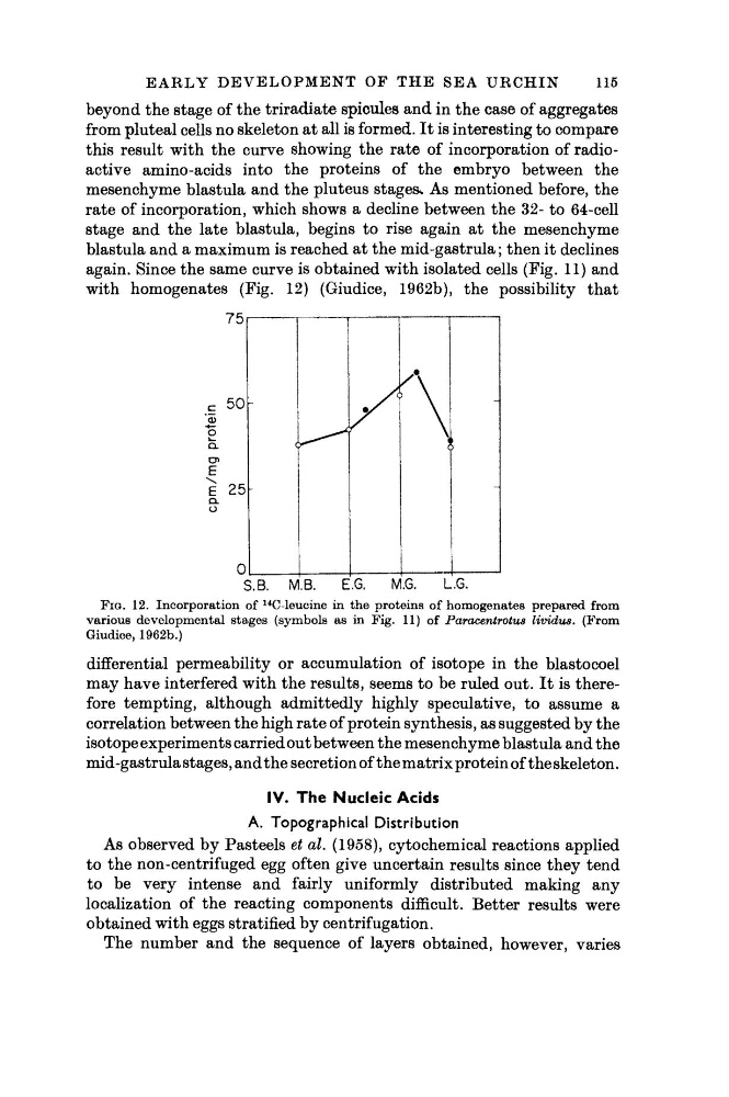
this result with the curve showing the rate of incorporation of radioactive amino-acids into the proteins of the embryo between the
mesenchyme blastula and the pluteus stages* As mentioned before, the
rate of incorporation, which shows a decline between the 32- to 64-cell
stage and the late blastula, begins to rise again at the mesenchyme
blastula and a maximum is reached at the mid-gastrula ; then it declines
again. Since the same curve is obtained with isolated cells (Fig. 11) and
with homogenates (Fig. 12) (Giudice, 1962b), the possibility that
75i
1
1
1
1
1
CL
01
ι
I
1
1 1
S.B.
M.B.
E.G.
M.G.
L.G.
FIG. 12. Incorporation of
14
C-leucine in the proteins of homogenates prepared from
various developmental stages (symbols as in Fig. 11) of Paracentrotus lividus. (From
Giudice, 1962b.)
differential permeability or accumulation of isotope in the blastocoel
may have interfered with the results, seems to be ruled out. It is therefore tempting, although admittedly highly speculative, to assume a
correlation between the high rate of protein synthesis, as suggested by the
isotope experiments carried out between the mesenchyme blastula and the
mid-gastrula stages, and the secretion of the matrix protein of the skeleton.
IV. The Nucleic Acids
A. Topographical Distribution
As observed by Pasteels et al. (1958), cytochemical reactions applied
to the non-centrifuged egg often give uncertain results since they tend
to be very intense and fairly uniformly distributed making any
localization of the reacting components difficult. Better results were
obtained with eggs stratified by centrifugation.
The number and the sequence of layers obtained, however, varies
115
beyond the stage of the triradiate spicules and in the case of aggregates
from pluteal cells no skeleton at all is formed. It is interesting to compare
this result with the curve showing the rate of incorporation of radioactive amino-acids into the proteins of the embryo between the
mesenchyme blastula and the pluteus stages* As mentioned before, the
rate of incorporation, which shows a decline between the 32- to 64-cell
stage and the late blastula, begins to rise again at the mesenchyme
blastula and a maximum is reached at the mid-gastrula ; then it declines
again. Since the same curve is obtained with isolated cells (Fig. 11) and
with homogenates (Fig. 12) (Giudice, 1962b), the possibility that
75i
1
1
1
1
1
CL
01
ι
I
1
1 1
S.B.
M.B.
E.G.
M.G.
L.G.
FIG. 12. Incorporation of
14
C-leucine in the proteins of homogenates prepared from
various developmental stages (symbols as in Fig. 11) of Paracentrotus lividus. (From
Giudice, 1962b.)
differential permeability or accumulation of isotope in the blastocoel
may have interfered with the results, seems to be ruled out. It is therefore tempting, although admittedly highly speculative, to assume a
correlation between the high rate of protein synthesis, as suggested by the
isotope experiments carried out between the mesenchyme blastula and the
mid-gastrula stages, and the secretion of the matrix protein of the skeleton.
IV. The Nucleic Acids
A. Topographical Distribution
As observed by Pasteels et al. (1958), cytochemical reactions applied
to the non-centrifuged egg often give uncertain results since they tend
to be very intense and fairly uniformly distributed making any
localization of the reacting components difficult. Better results were
obtained with eggs stratified by centrifugation.
The number and the sequence of layers obtained, however, varies