

642
I L S E
L A S N I T Z K I
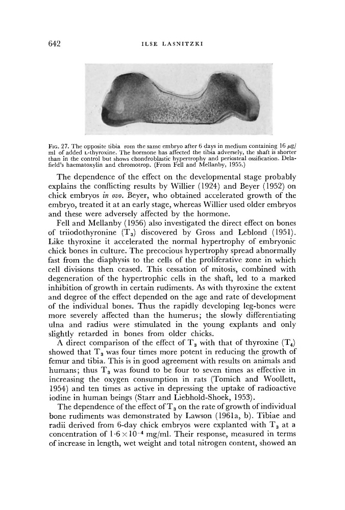
FIG. 27. The opposite tibia rom the same embryo after 6 days in medium containing 16 fig/
ml of added L-thyroxine. The hormone has affected the tibia adversely, the shaft is shorter
than in the control but shows chondroblastic hypertrophy and periosteal ossification. Delafield's haematoxylin and chromotrop. (From Fell and Mellanby, 1955.)
The dependence of the effect on the developmental stage probably
explains the conflicting results by Willier (1924) and Beyer (1952) on
chick embryos in ovo. Beyer, who obtained accelerated growth of the
embryo, treated it at an early stage, whereas Willier used older embryos
and these were adversely affected by the hormone.
Fell and Mellanby (1956) also investigated the direct effect on bones
of triiodothyronine ( T 3) discovered by Gross and Leblond (1951).
Like thyroxine it accelerated the normal hypertrophy of embryonic
chick bones in culture. The precocious hypertrophy spread abnormally
fast from the diaphysis to the cells of the proliferative zone in which
cell divisions then ceased. This cessation of mitosis, combined with
degeneration of the hypertrophic cells in the shaft, led to a marked
inhibition of growth in certain rudiments. As with thyroxine the extent
and degree of the effect depended on the age and rate of development
of the individual bones. Thus the rapidly developing leg-bones were
more severely affected than the humerus; the slowly differentiating
ulna and radius were stimulated in the young explants and only
slightly retarded in bones from older chicks.
A direct comparison of the effect of T 3 with that of thyroxine ( T 4)
showed that T 3 was four times more potent in reducing the growth of
femur and tibia. This is in good agreement with results on animals and
humans; thus T 3 was found to be four to seven times as effective in
increasing the oxygen consumption in rats (Tomich and Woollett,
1954) and ten times as active in depressing the uptake of radioactive
iodine in human beings (Starr and Liebhold-Shoek, 1953).
The dependence ofthe effect of T 3 on the rate of growth of individual
bone rudiments was demonstrated by Lawson (1961a, b ) . Tibiae and
radii derived from 6-day chick embryos were explanted with T 3 at a
concentration of 1-6 X 10~4 mg/ml. Their response, measured in terms
of increase in length, wet weight and total nitrogen content, showed an