

16. A C T I O N S OF H O R M O N E S
641
increased rate of proliferation in the cartilage and evidence for hastened
maturity in the matrix. Results on the effects of thyroxine on the development of chick bones in ovo have been conflicting. Willier (1924)
found that grafting of thyroid into 7-10-day chick embryos considerably
inhibited the growth of limb bones while Beyer (1952) obtained
increases in the wet and dry weights of chick embryos from eggs
injected with thyroxine before incubation.
Studies by Fell and Mellanby (1955) on the effects of thyroxine on
organ cultures of bone rudiments showed that the hormone had a
direct effect on the tissue. Chicklong-bone rudiments were treated at
different stages of development with a concentration of thyroxine of
the same order as that present in hyperthyroidal human serum. The
hormone accelerated the normal hypertrophy of the cartilage cells but
also caused regressive changes. The same concentration was stimulatory or harmful, depending on the age and growth rate of the particular bone used. Young rudiments were more easily stimulated than
the older bones; of all the bones tested the humerus responded best at
all ages and showed acceleration of hypertrophy leading to a rapid
extension of the bone and temporary increase in size over that of the
controls (see V o l . 2, Chapter 4 ) . The radius which differentiates more
slowly than the other bones was occasionally stimulated at the precartilaginous stage but always in the older explants. In the tibia and femur the
maturation of the cartilagewas some times accelerated in young bones
but only slightly and temporarily at the older stage.
The older bones were more susceptible to the toxic action of the
hormone which caused a degeneration of the cartilage cells. Treatment
with higher doses than that present in hyperthyroidal serum demonstrated a great difference in susceptibility between the various rudiments.
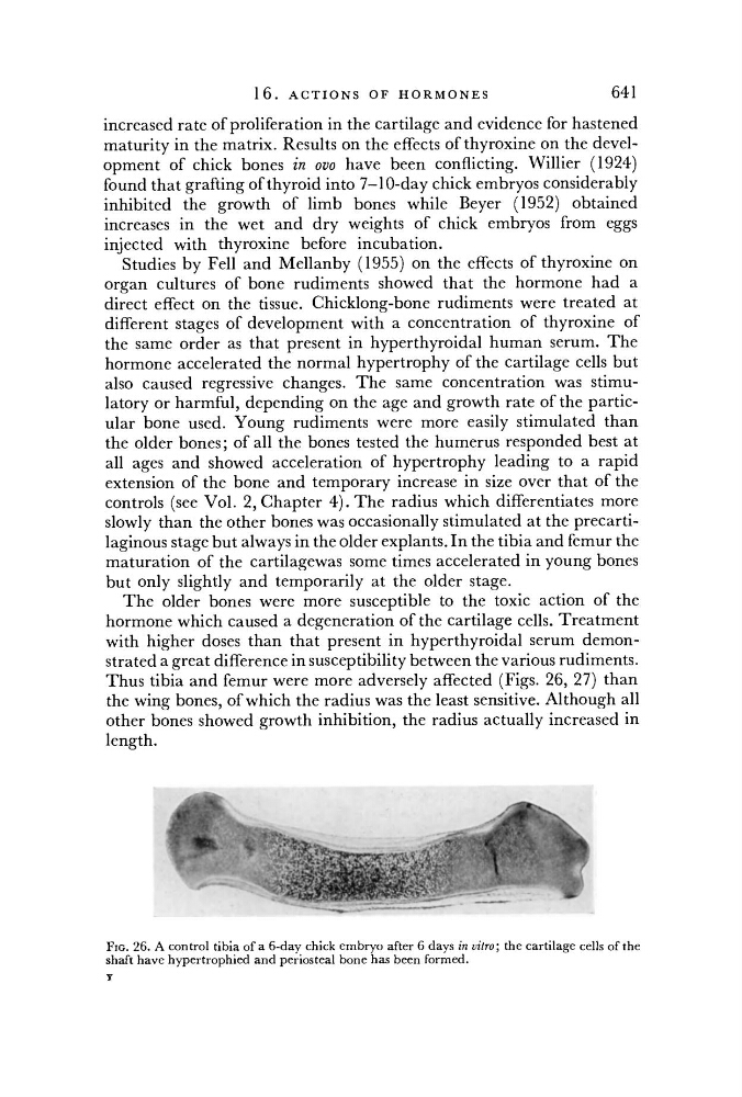
Thus tibia and femur were more adversely affected (Figs. 26, 27) than
the wing bones, of which the radius was the least sensitive. Although all
other bones showed growth inhibition, the radius actually increased in
length.
FIG. 26. A control tibia of a 6-day chick embryo after 6 days in vitro; the cartilage cells of the
shaft have hypertrophied and periosteal bone has been formed.
Y