

628
I L S E
L A S N I T Z K I
four times the control size, but higher doses were necessary to promote
the growth of the connective tissue of the tunica propria and of the
epithelium which occasionally became multilayered.
The role of the sex hormones in the causation of prostatic tumours
is now widely recognized. Both the benign enlargement of the gland
and prostatic cancer have been attributed to a disturbance of the
androgen-oestrogen balance and in particular to a relative increase of
the oestrogen level during advancing age. The question arises whether
the hormonal changes are a primary cause or whether mature cells
respond in a different manner from young cells to alterations in their
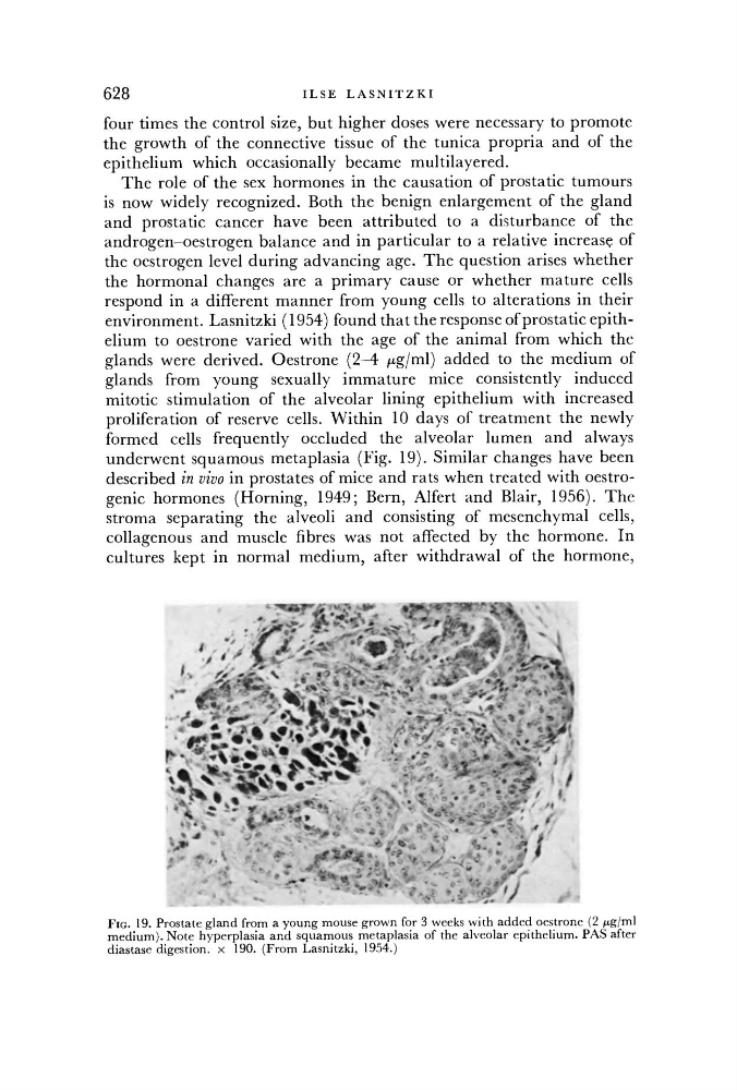
environment. Lasnitzki (1954) found that the response of prostatic epithelium to oestrone varied with the age of the animal from which the
glands were derived. Oestrone (2-4 /xg/ml) added to the medium of
glands from young sexually immature mice consistently induced
mitotic stimulation of the alveolar lining epithelium with increased
proliferation of reserve cells. Within 10 days of treatment the newly
formed cells frequently occluded the alveolar lumen and always
underwent squamous metaplasia (Fig. 19). Similar changes have been
described in vivo in prostates of mice and rats when treated with oestrogenic hormones (Horning, 1949; Bern, Alfert and Blair, 1956). The
stroma separating the alveoli and consisting of mesenchymal cells,
collagenous and muscle fibres was not affected by the hormone. In
cultures kept in normal medium, after withdrawal of the hormone,
FIG. 19. Prostate gland from a young mouse grown for 3 weeks with added oestrone (2 /ng/ml
medium). Note hyperplasia and squamous metaplasia of the alveolar epithelium. PAS after
diastase digestion, x 190. (From Lasnitzki, 1954.)