

618
I L S E
L A S N I T Z K I
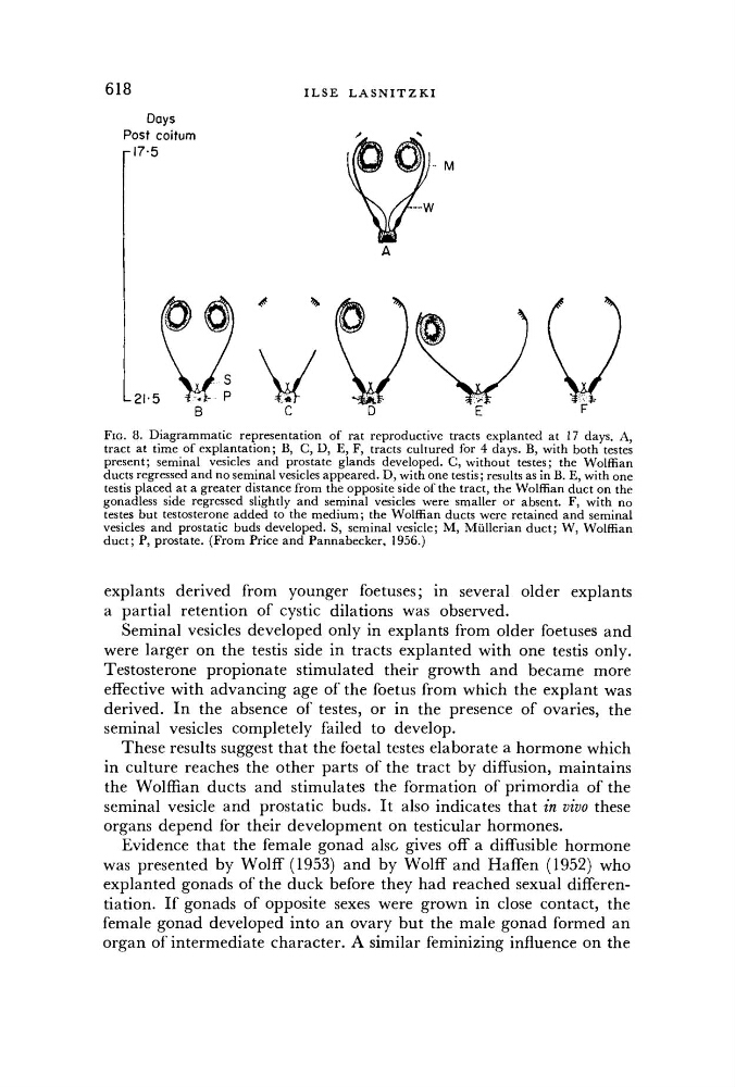
Days
FIG. 8. Diagrammatic representation of rat reproductive tracts explanted at 17 days. A,
tract at time of explantation; B, C, D, E, F, tracts cultured for 4 days. B, with both testes
present; seminal vesicles and prostate glands developed. G, without testes; the Wolffian
ducts regressed and no seminal vesicles appeared. D , with one testis; results as in B. E, with one
testis placed at a greater distance from the opposite side of the tract, the Wolffian duct on the
gonadless side regressed slightly and seminal vesicles were smaller or absent. F, with no
testes but testosterone added to the medium; the Wolffian ducts were retained and seminal
vesicles and prostatic buds developed. S, seminal vesicle; M , Miillerian duct; W , Wolffian
duct; P, prostate. (From Price and Pannabecker, 1956.)
explants derived from younger foetuses; in several older explants
a partial retention of cystic dilations was observed.
Seminal vesicles developed only in explants from older foetuses and
were larger on the testis side in tracts explanted with one testis only.
Testosterone propionate stimulated their growth and became more
effective with advancing age of the foetus from which the explant was
derived. In the absence of testes, or in the presence of ovaries, the
seminal vesicles completely failed to develop.
These results suggest that the foetal testes elaborate a hormone which
in culture reaches the other parts of the tract by diffusion, maintains
the Wolffian ducts and stimulates the formation of primordia of the
seminal vesicle and prostatic buds. It also indicates that in vivo these
organs depend for their development on testicular hormones.
Evidence that the female gonad also gives off a diffusible hormone
was presented by Wolff (1953) and by Wolff and Haffen (1952) who
explanted gonads of the duck before they had reached sexual differentiation. If gonads of opposite sexes were grown in close contact, the
female gonad developed into an ovary but the male gonad formed an
organ of intermediate character. A similar feminizing influence on the