

326
W. B. SCHAEFER
chromogenic at room temperature of 25°C. It slowly hydrolyses Tween-SO,
gives a positive arylsulphatase test after three to five days, and reduces
nitrate. Since the original publication on seven strains, 13 more isolates
have been found in the U.S.A. (Schaefer, unpublished data).
D. Mycobacterium avium and Mycobacterium intracellulare
M. avium was described in 1890 by Maffucci as the causative agent of
avian tuberculosis. M. intracellulare is indistinguishable from M . avium in
its cultural and biochemical properties, but does not cause tuberculosis
in birds. Only in the early 1950s was it noted by Smith and Stergus (1964)
at the Battey State Hospital as the apparent cause of pulmonary tuberculosis
in man. It was called the “Battey” bacillus. The name was changed to
M. intracellulare (Runyon, 1967).
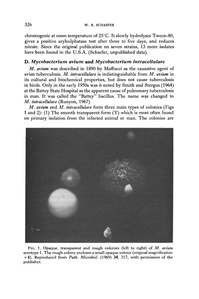
M . avium and M. intracellulare form three main types of colonies (Figs
1 and 2): (1) The smooth transparent form (T) which is most often found
on primary isolation from the infected animal or man. The colonies are
FIG. 1. Opaque, transparent and rough colonies (left to right) of M. avium
serotype 1. The rough colony encloses a small opaque colony (original magnification
x 8). Reproduced from Path. Microbiol. (1969) 34, 317, with permission of the
publisher.
W. B. SCHAEFER
chromogenic at room temperature of 25°C. It slowly hydrolyses Tween-SO,
gives a positive arylsulphatase test after three to five days, and reduces
nitrate. Since the original publication on seven strains, 13 more isolates
have been found in the U.S.A. (Schaefer, unpublished data).
D. Mycobacterium avium and Mycobacterium intracellulare
M. avium was described in 1890 by Maffucci as the causative agent of
avian tuberculosis. M. intracellulare is indistinguishable from M . avium in
its cultural and biochemical properties, but does not cause tuberculosis
in birds. Only in the early 1950s was it noted by Smith and Stergus (1964)
at the Battey State Hospital as the apparent cause of pulmonary tuberculosis
in man. It was called the “Battey” bacillus. The name was changed to
M. intracellulare (Runyon, 1967).
M . avium and M. intracellulare form three main types of colonies (Figs
1 and 2): (1) The smooth transparent form (T) which is most often found
on primary isolation from the infected animal or man. The colonies are
FIG. 1. Opaque, transparent and rough colonies (left to right) of M. avium
serotype 1. The rough colony encloses a small opaque colony (original magnification
x 8). Reproduced from Path. Microbiol. (1969) 34, 317, with permission of the
publisher.