

112
A. SARAGEA ET AL.
growth is treated for 2 h by chloroform vapour. Young liquid cultures
(2-3 h incubation at 37” in cystinated Pope Linggood broth) of the strains
to be tested are then inoeulated with a thin loop, at right angles to the
killed bacteriocinogenic growth. The streaks are made on both sides of the
diametrical band, from the edges of the bacteriocinogenic culture to the
margins of the plate (Fig. 22).
Plates of 12 cm diameter accommodate 16-20 strains to be tested. The
results are recorded after overnight incubation at 37°C. The growth of
a bacteriocin sensitive strain is inhibited over a smaller or bigger area
starting from the central bacteriocinogenic band. The whole range of
reactions is recorded. The minimum positive inhibition area is 2 mm, with
or without isolated resistant colonies.
Smaller inhibition zones than 2 mm are considered only when the strain
would not otherwise be defined.
(d) Media used. The medium recommended for bacteriocin typing of
C. diphtheriae is brain-heart infusion with 1.3% agar, 0.001% cystine to
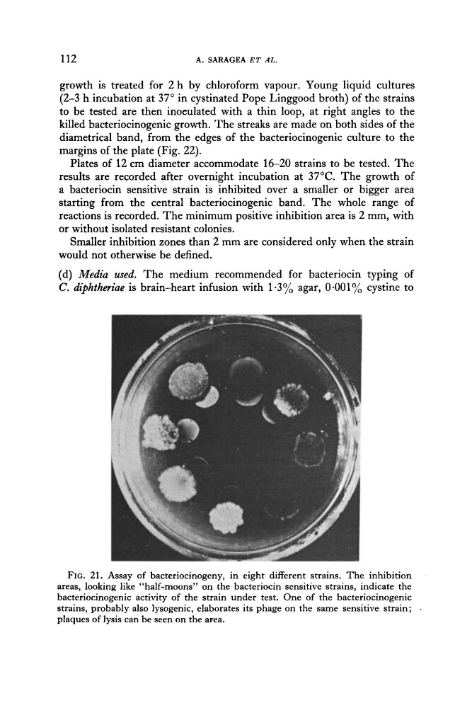
FIG. 21. Assay of bacteriocinogeny, in eight different strains. The inhibition
areas, looking like “half-moons” on the bacteriocin sensitive strains, indicate the
bacteriocinogenic activity of the strain under test. One of the bacteriocinogenic
strains, probably also lysogenic, elaborates its phage on the same sensitive strain;
plaques of lysis can be seen on the area.
A. SARAGEA ET AL.
growth is treated for 2 h by chloroform vapour. Young liquid cultures
(2-3 h incubation at 37” in cystinated Pope Linggood broth) of the strains
to be tested are then inoeulated with a thin loop, at right angles to the
killed bacteriocinogenic growth. The streaks are made on both sides of the
diametrical band, from the edges of the bacteriocinogenic culture to the
margins of the plate (Fig. 22).
Plates of 12 cm diameter accommodate 16-20 strains to be tested. The
results are recorded after overnight incubation at 37°C. The growth of
a bacteriocin sensitive strain is inhibited over a smaller or bigger area
starting from the central bacteriocinogenic band. The whole range of
reactions is recorded. The minimum positive inhibition area is 2 mm, with
or without isolated resistant colonies.
Smaller inhibition zones than 2 mm are considered only when the strain
would not otherwise be defined.
(d) Media used. The medium recommended for bacteriocin typing of
C. diphtheriae is brain-heart infusion with 1.3% agar, 0.001% cystine to
FIG. 21. Assay of bacteriocinogeny, in eight different strains. The inhibition
areas, looking like “half-moons” on the bacteriocin sensitive strains, indicate the
bacteriocinogenic activity of the strain under test. One of the bacteriocinogenic
strains, probably also lysogenic, elaborates its phage on the same sensitive strain;
plaques of lysis can be seen on the area.