

98
3. Benzenoids
Australian workers isolated a mixture of pigments from the spines and tests
of the echinoid Salmacis sphaeroides, and by comparison with literature data
surmised that one of their pigments (which was not adsorbed on calcium
carbonate) might be identical with a substance referred to as spinochrome Β
and for which several incorrect structures had been proposed. With the aid
of nmr data Gough and Sutherland (1964) proved their pigment to have the
structure 2,3,7-trihydroxyjuglone (18), a structure that had been assigned to
spinochrome Ν by Kuroda and that had been proved by synthesis (Smith and
Thomson, 1961). By direct comparison of all available samples Gough and
Sutherland (1964) showed that their pigment 18 was identical with no fewer
than six other sea urchin pigments that had been described in the literature
under various names, and some of them with insufficient characterization.
A decisive turning point in sea urchin pigment research came about by
Chang's (1964a) use of nmr techniques and by his systematic and successful
search for an adequate purification procedure. The traditional chromatography introduced by Lederer and Glaser (1938), was carried out on calcium
(occasionally magnesium) carbonate. This adsorbent parallels the natural
substrate of the pigments but generally does not allow elution by solvent to
take place. Instead, partially developed columns are cut, redissolved, and the
process is repeated. Chang (1964a) after an extensive study found that silica
gel that was deactivated by prior acid treatment permitted separation of the
pigments and elution by solvents.
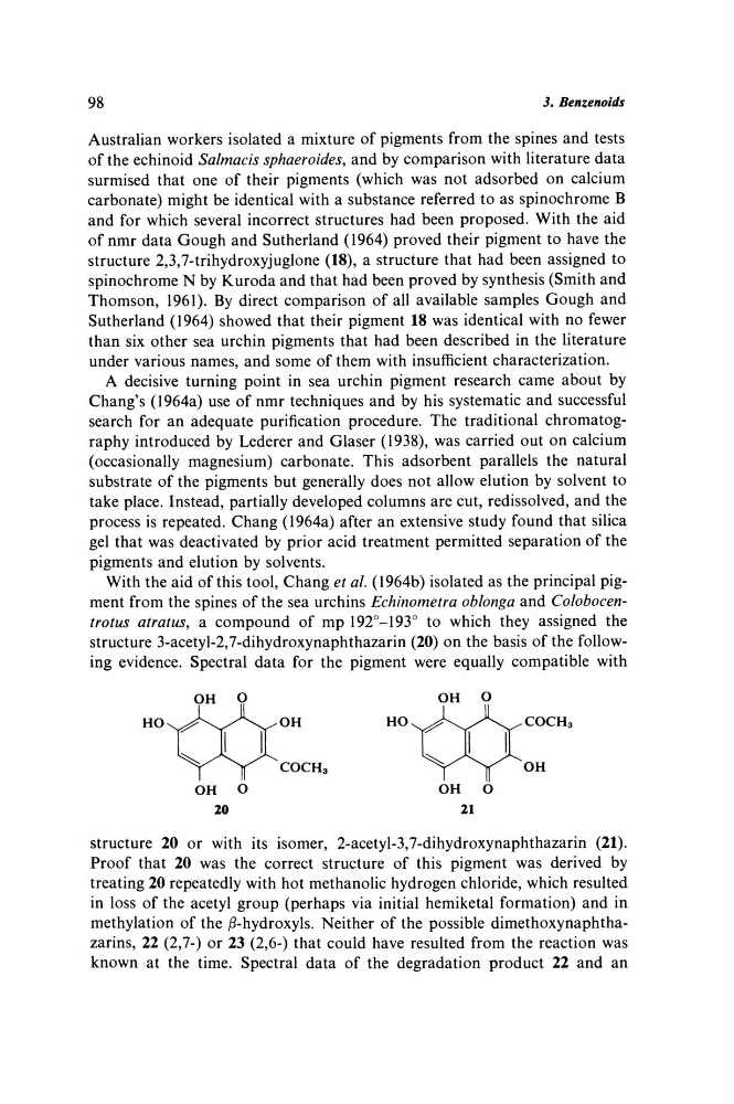
With the aid of this tool, Chang et al. (1964b) isolated as the principal pigment from the spines of the sea urchins Echinometra oblonga and Colobocentrotus atratus, a compound of mp 192°-193° to which they assigned the
structure 3-acetyl-2,7-dihydroxynaphthazarin (20) on the basis of the following evidence. Spectral data for the pigment were equally compatible with
OH
Ο
OH
Ο
HO
V^^rr"
OH
Ho^^k^^A.
COCH 3
OH
Ο
OH
Ο
20
21
structure 20 or with its isomer, 2-acetyl-3,7-dihydroxynaphthazarin (21).
Proof that 20 was the correct structure of this pigment was derived by
treating 20 repeatedly with hot methanolic hydrogen chloride, which resulted
in loss of the acetyl group (perhaps via initial hemiketal formation) and in
methylation of the ß-hydroxyls. Neither of the possible dimethoxynaphthazarins, 22 (2,7-) or 23 (2,6-) that could have resulted from the reaction was
known at the time. Spectral data of the degradation product 22 and an
3. Benzenoids
Australian workers isolated a mixture of pigments from the spines and tests
of the echinoid Salmacis sphaeroides, and by comparison with literature data
surmised that one of their pigments (which was not adsorbed on calcium
carbonate) might be identical with a substance referred to as spinochrome Β
and for which several incorrect structures had been proposed. With the aid
of nmr data Gough and Sutherland (1964) proved their pigment to have the
structure 2,3,7-trihydroxyjuglone (18), a structure that had been assigned to
spinochrome Ν by Kuroda and that had been proved by synthesis (Smith and
Thomson, 1961). By direct comparison of all available samples Gough and
Sutherland (1964) showed that their pigment 18 was identical with no fewer
than six other sea urchin pigments that had been described in the literature
under various names, and some of them with insufficient characterization.
A decisive turning point in sea urchin pigment research came about by
Chang's (1964a) use of nmr techniques and by his systematic and successful
search for an adequate purification procedure. The traditional chromatography introduced by Lederer and Glaser (1938), was carried out on calcium
(occasionally magnesium) carbonate. This adsorbent parallels the natural
substrate of the pigments but generally does not allow elution by solvent to
take place. Instead, partially developed columns are cut, redissolved, and the
process is repeated. Chang (1964a) after an extensive study found that silica
gel that was deactivated by prior acid treatment permitted separation of the
pigments and elution by solvents.
With the aid of this tool, Chang et al. (1964b) isolated as the principal pigment from the spines of the sea urchins Echinometra oblonga and Colobocentrotus atratus, a compound of mp 192°-193° to which they assigned the
structure 3-acetyl-2,7-dihydroxynaphthazarin (20) on the basis of the following evidence. Spectral data for the pigment were equally compatible with
OH
Ο
OH
Ο
HO
V^^rr"
OH
Ho^^k^^A.
COCH 3
OH
Ο
OH
Ο
20
21
structure 20 or with its isomer, 2-acetyl-3,7-dihydroxynaphthazarin (21).
Proof that 20 was the correct structure of this pigment was derived by
treating 20 repeatedly with hot methanolic hydrogen chloride, which resulted
in loss of the acetyl group (perhaps via initial hemiketal formation) and in
methylation of the ß-hydroxyls. Neither of the possible dimethoxynaphthazarins, 22 (2,7-) or 23 (2,6-) that could have resulted from the reaction was
known at the time. Spectral data of the degradation product 22 and an