

Biochemical Systematics and Ecology 96 (2021) 104262
7
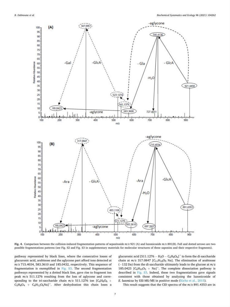
pathway represented by black lines, where the consecutive losses of
glucuronic acid, arabinose and the aglycone part afford ions detected at
m/z 715.4034, 583.3610 and 185.0432, respectively. This sequence of
fragmentation is exemplified in Fig. S3. The second fragmentation
pathways represented by a dotted black line, gave rise to fragment ion
peak m/z 511.1276 resulting from the loss of aglycone and corresponding to the tri-saccharide chain m/z 511.1276 ion [C 6 H 8 O 6 +
C 5 H 8 O 4 + C 6 H 12 O 6 Na]
+
. After deshydration this chain loses a
glucuronic acid [511.1276 − H 2 O − C 6 H 8 O 6 ]
+
to form the di-saccharide
chain at m/z 317.0847 [C 11 H 18 O 9 Na]. The elimination of arabinose
(− 132 Da) from the di-saccharide ultimately leads to the glucose at m/z
185.0423 [C 6 H 10 O 5 + Na]
+
. The complete dissociation pathway is
described in Fig. S3. Indeed, these two fragmentations gave signals
consistent with those obtained by analyzing the luzonicoside of
E. luzonicus by ESI-MS/MS in positive mode (Kicha et al., 2015).
This result suggests that the CID spectra of the m/z 891.4353 are in
Fig. 4. Comparison between the collision-induced fragmentation patterns of sepositoside m/z 921 (A) and luzonicoside m/z 891(B). Full and dotted arrows are two
possible fragmentations patterns (see Fig. S2 and Fig. S3 in supplementary materials for molecular structures of these saponins and their respective fragments).
B. Dahmoune et al.
7
pathway represented by black lines, where the consecutive losses of
glucuronic acid, arabinose and the aglycone part afford ions detected at
m/z 715.4034, 583.3610 and 185.0432, respectively. This sequence of
fragmentation is exemplified in Fig. S3. The second fragmentation
pathways represented by a dotted black line, gave rise to fragment ion
peak m/z 511.1276 resulting from the loss of aglycone and corresponding to the tri-saccharide chain m/z 511.1276 ion [C 6 H 8 O 6 +
C 5 H 8 O 4 + C 6 H 12 O 6 Na]
+
. After deshydration this chain loses a
glucuronic acid [511.1276 − H 2 O − C 6 H 8 O 6 ]
+
to form the di-saccharide
chain at m/z 317.0847 [C 11 H 18 O 9 Na]. The elimination of arabinose
(− 132 Da) from the di-saccharide ultimately leads to the glucose at m/z
185.0423 [C 6 H 10 O 5 + Na]
+
. The complete dissociation pathway is
described in Fig. S3. Indeed, these two fragmentations gave signals
consistent with those obtained by analyzing the luzonicoside of
E. luzonicus by ESI-MS/MS in positive mode (Kicha et al., 2015).
This result suggests that the CID spectra of the m/z 891.4353 are in
Fig. 4. Comparison between the collision-induced fragmentation patterns of sepositoside m/z 921 (A) and luzonicoside m/z 891(B). Full and dotted arrows are two
possible fragmentations patterns (see Fig. S2 and Fig. S3 in supplementary materials for molecular structures of these saponins and their respective fragments).
B. Dahmoune et al.