

Aiken
46
acids are present in more complex, proteinaceous forms requiring hydrolysis (Cowie and
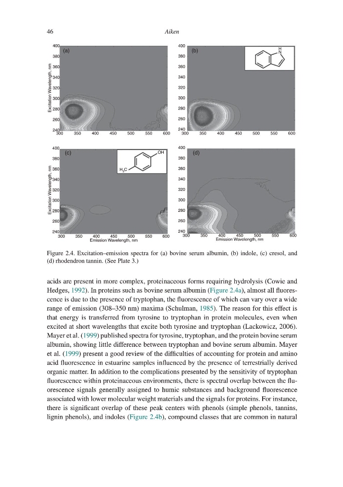
Hedges, 1992). In proteins such as bovine serum albumin (Figure 2.4a), almost all fluorescence is due to the presence of tryptophan, the fluorescence of which can vary over a wide
range of emission (308–350 nm) maxima (Schulman, 1985). The reason for this effect is
that energy is transferred from tyrosine to tryptophan in protein molecules, even when
excited at short wavelengths that excite both tyrosine and tryptophan (Lackowicz, 2006).
Mayer et al. (1999) published spectra for tyrosine, tryptophan, and the protein bovine serum
albumin, showing little difference between tryptophan and bovine serum albumin. Mayer
et al. (1999) present a good review of the difficulties of accounting for protein and amino
acid fluorescence in estuarine samples influenced by the presence of terrestrially derived
organic matter. In addition to the complications presented by the sensitivity of tryptophan
fluorescence within proteinaceous environments, there is spectral overlap between the fluorescence signals generally assigned to humic substances and background fluorescence
associated with lower molecular weight materials and the signals for proteins. For instance,
there is significant overlap of these peak centers with phenols (simple phenols, tannins,
lignin phenols), and indoles (Figure 2.4b), compound classes that are common in natural
400
380
Excitation Wavelength, nm
360
340
320
300
280
260
300
350
400
450
500
550
600
300
350
400
450
Emission Wavelength, nm
500
550
600
300
350
400
450
Emission Wavelength, nm
500
550
600
300
350
400
450
500
550
600
240
400
380
Excitation Wavelength, nm
360
340
320
300
280
260
240
400
380
360
340
320
300
280
260
240
400
H
N
OH
H 3 C
380
360
340
320
300
280
260
240
(a)
(b)
(d)
(c)
Figure 2.4. Excitation–emission spectra for (a) bovine serum albumin, (b) indole, (c) cresol, and
(d) rhodendron tannin. (See Plate 3.)
46
acids are present in more complex, proteinaceous forms requiring hydrolysis (Cowie and
Hedges, 1992). In proteins such as bovine serum albumin (Figure 2.4a), almost all fluorescence is due to the presence of tryptophan, the fluorescence of which can vary over a wide
range of emission (308–350 nm) maxima (Schulman, 1985). The reason for this effect is
that energy is transferred from tyrosine to tryptophan in protein molecules, even when
excited at short wavelengths that excite both tyrosine and tryptophan (Lackowicz, 2006).
Mayer et al. (1999) published spectra for tyrosine, tryptophan, and the protein bovine serum
albumin, showing little difference between tryptophan and bovine serum albumin. Mayer
et al. (1999) present a good review of the difficulties of accounting for protein and amino
acid fluorescence in estuarine samples influenced by the presence of terrestrially derived
organic matter. In addition to the complications presented by the sensitivity of tryptophan
fluorescence within proteinaceous environments, there is spectral overlap between the fluorescence signals generally assigned to humic substances and background fluorescence
associated with lower molecular weight materials and the signals for proteins. For instance,
there is significant overlap of these peak centers with phenols (simple phenols, tannins,
lignin phenols), and indoles (Figure 2.4b), compound classes that are common in natural
400
380
Excitation Wavelength, nm
360
340
320
300
280
260
300
350
400
450
500
550
600
300
350
400
450
Emission Wavelength, nm
500
550
600
300
350
400
450
Emission Wavelength, nm
500
550
600
300
350
400
450
500
550
600
240
400
380
Excitation Wavelength, nm
360
340
320
300
280
260
240
400
380
360
340
320
300
280
260
240
400
H
N
OH
H 3 C
380
360
340
320
300
280
260
240
(a)
(b)
(d)
(c)
Figure 2.4. Excitation–emission spectra for (a) bovine serum albumin, (b) indole, (c) cresol, and
(d) rhodendron tannin. (See Plate 3.)