

Osburn, Del Vecchio, and Boyd
250
(e.g., flavenoids) having extended π-conjugation. The molecular weight separation dramatically modified SF features. On adding Al(III) concentrations at 4–400 μM, SF increased
for all three peaks identified and most strongly for peak III, which was most prevalent in the
500–1000 MW fraction. Titration curves eventually reached a saturation point and these
were interpreted to suggest that deprotonated (at pH 3.5) carboxyl–Al complexes were
inducing fluorescence.
Ryan et al. (1996), Elkins and Nelson (2001, 2002), and Zhao and Nelson (2005) all
found that Al(III) strongly interacts with salicylic acid moieties in fulvic acids, and that
Al(III)–salicylic acid complexes produced new and highly intense fluorescent peaks in
EEMs. Salicyclic acid has both phenolic and carboxylic functionality and is a major component of FA. A common spectral change observed by these workers was a significant
20 nm red shift in the excitation peak maxima and a 20 nm blue shift in emission peak
maxima, after addition of Al(III) to FA solutions. The same effect occurred for solutions
of salicyclic acid complexed with Al(III) (Elkins and Nelson, 2001). However, Zhao and
Nelson (2005) also observed that although Al(III) outcompeted other metals for available
binding sites on FA and formed a new fluorescent complex with FA, Fe(III) complexation
by FA actually quenched the new Al(III)–FA fluorescence. During complexation, metals
bind to anionic functional groups via donation of electrons in a binding environment that
may range from ionic to covalent. Zhao and Nelson (2005) noted that the uniquely low
covalent and high ionic properties of ions such as Al(III) and Fe(III) alter the electronic
structure of FA through complex formation and inducing these contrasting effects on FA
fluorescence.
0
20
40
60
80
100
120
140
0
0.00005 0.0001 0.00015 0.0002
[M n+ ] (M)
Relative Fluorescence Intensity
Al(III); Em = 424 nm
Cd(II); Em = 448 nm
Cu(II); Em = 448 nm
pH = 5; Ex = 344nm
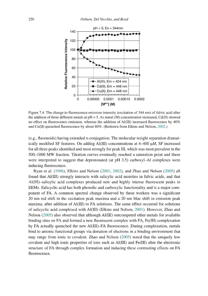
Figure 7.4. The change to fluorescence emission intensity (excitation of 344 nm) of fulvic acid after
the addition of three different metals at pH = 5. As metal (M) concentration increased, Cd(II) showed
no effect on fluorescence emission, whereas the addition of Al(III) increased fluorescence by 40%
and Cu(II) quenched fluorescence by about 60%. (Redrawn from Elkins and Nelson, 2002.)
250
(e.g., flavenoids) having extended π-conjugation. The molecular weight separation dramatically modified SF features. On adding Al(III) concentrations at 4–400 μM, SF increased
for all three peaks identified and most strongly for peak III, which was most prevalent in the
500–1000 MW fraction. Titration curves eventually reached a saturation point and these
were interpreted to suggest that deprotonated (at pH 3.5) carboxyl–Al complexes were
inducing fluorescence.
Ryan et al. (1996), Elkins and Nelson (2001, 2002), and Zhao and Nelson (2005) all
found that Al(III) strongly interacts with salicylic acid moieties in fulvic acids, and that
Al(III)–salicylic acid complexes produced new and highly intense fluorescent peaks in
EEMs. Salicyclic acid has both phenolic and carboxylic functionality and is a major component of FA. A common spectral change observed by these workers was a significant
20 nm red shift in the excitation peak maxima and a 20 nm blue shift in emission peak
maxima, after addition of Al(III) to FA solutions. The same effect occurred for solutions
of salicyclic acid complexed with Al(III) (Elkins and Nelson, 2001). However, Zhao and
Nelson (2005) also observed that although Al(III) outcompeted other metals for available
binding sites on FA and formed a new fluorescent complex with FA, Fe(III) complexation
by FA actually quenched the new Al(III)–FA fluorescence. During complexation, metals
bind to anionic functional groups via donation of electrons in a binding environment that
may range from ionic to covalent. Zhao and Nelson (2005) noted that the uniquely low
covalent and high ionic properties of ions such as Al(III) and Fe(III) alter the electronic
structure of FA through complex formation and inducing these contrasting effects on FA
fluorescence.
0
20
40
60
80
100
120
140
0
0.00005 0.0001 0.00015 0.0002
[M n+ ] (M)
Relative Fluorescence Intensity
Al(III); Em = 424 nm
Cd(II); Em = 448 nm
Cu(II); Em = 448 nm
pH = 5; Ex = 344nm
Figure 7.4. The change to fluorescence emission intensity (excitation of 344 nm) of fulvic acid after
the addition of three different metals at pH = 5. As metal (M) concentration increased, Cd(II) showed
no effect on fluorescence emission, whereas the addition of Al(III) increased fluorescence by 40%
and Cu(II) quenched fluorescence by about 60%. (Redrawn from Elkins and Nelson, 2002.)