

Osburn, Del Vecchio, and Boyd
246
and protein fluorescence. Proteinaceous fluorescence emission intensities were observed
to either show no variations with pH (Spencer et al., 2007b) or increasing signal with
increasing pH (Baker et al., 2007). Several explanations have been given for the marked
variability in results with proteinaceous materials. They are thought to be differentially
impacted by colloidal material that may protect moieties from solution exposure, change
tertiary structure in response to protonation–deprotonation, and perhaps change based on
their acid–base content (Baker et al., 2007).
no Cu
Cu
0
0
2
4
6
8
10
12
10
20
30
40
50
60
70
80
90
100
pH
0
2
4
6
8
pH
Relative fluorescence
(a)
0
10
20
30
40
50
60
70
80
90
100
Relative fluorescence
(b)
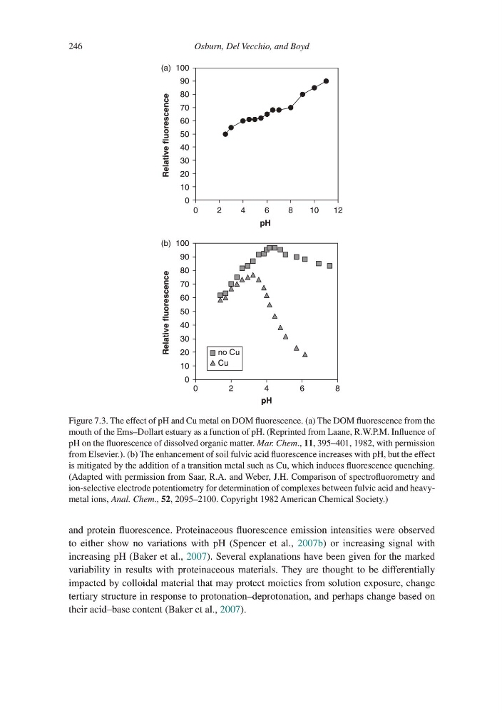
Figure 7.3. The effect of pH and Cu metal on DOM fluorescence. (a) The DOM fluorescence from the
mouth of the Ems–Dollart estuary as a function of pH. (Reprinted from Laane, R.W.P.M. Influence of
pH on the fluorescence of dissolved organic matter. Mar. Chem., 11, 395–401, 1982, with permission
from Elsevier.). (b) The enhancement of soil fulvic acid fluorescence increases with pH, but the effect
is mitigated by the addition of a transition metal such as Cu, which induces fluorescence quenching.
(Adapted with permission from Saar, R.A. and Weber, J.H. Comparison of spectrofluorometry and
ion-selective electrode potentiometry for determination of complexes between fulvic acid and heavymetal ions, Anal. Chem., 52, 2095–2100. Copyright 1982 American Chemical Society.)
246
and protein fluorescence. Proteinaceous fluorescence emission intensities were observed
to either show no variations with pH (Spencer et al., 2007b) or increasing signal with
increasing pH (Baker et al., 2007). Several explanations have been given for the marked
variability in results with proteinaceous materials. They are thought to be differentially
impacted by colloidal material that may protect moieties from solution exposure, change
tertiary structure in response to protonation–deprotonation, and perhaps change based on
their acid–base content (Baker et al., 2007).
no Cu
Cu
0
0
2
4
6
8
10
12
10
20
30
40
50
60
70
80
90
100
pH
0
2
4
6
8
pH
Relative fluorescence
(a)
0
10
20
30
40
50
60
70
80
90
100
Relative fluorescence
(b)
Figure 7.3. The effect of pH and Cu metal on DOM fluorescence. (a) The DOM fluorescence from the
mouth of the Ems–Dollart estuary as a function of pH. (Reprinted from Laane, R.W.P.M. Influence of
pH on the fluorescence of dissolved organic matter. Mar. Chem., 11, 395–401, 1982, with permission
from Elsevier.). (b) The enhancement of soil fulvic acid fluorescence increases with pH, but the effect
is mitigated by the addition of a transition metal such as Cu, which induces fluorescence quenching.
(Adapted with permission from Saar, R.A. and Weber, J.H. Comparison of spectrofluorometry and
ion-selective electrode potentiometry for determination of complexes between fulvic acid and heavymetal ions, Anal. Chem., 52, 2095–2100. Copyright 1982 American Chemical Society.)