

Gilchrist and Reynolds
158
5.3.2 Basic Spectrofluorimeter Design
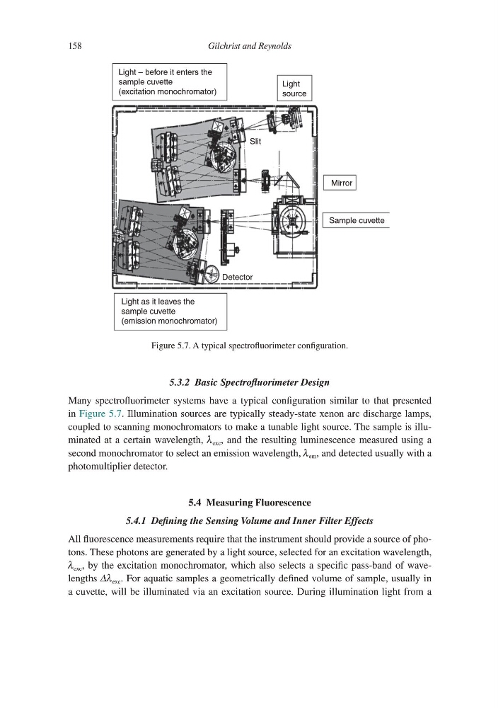
Many spectrofluorimeter systems have a typical configuration similar to that presented
in Figure 5.7. Illumination sources are typically steady-state xenon arc discharge lamps,
coupled to scanning monochromators to make a tunable light source. The sample is illuminated at a certain wavelength, λ exc , and the resulting luminescence measured using a
second monochromator to select an emission wavelength, λ em , and detected usually with a
photomultiplier detector.
5.4 Measuring Fluorescence
5.4.1 Defining the Sensing Volume and Inner Filter Effects
All fluorescence measurements require that the instrument should provide a source of photons. These photons are generated by a light source, selected for an excitation wavelength,
λ exc , by the excitation monochromator, which also selects a specific pass-band of wavelengths Δλ exc . For aquatic samples a geometrically defined volume of sample, usually in
a cuvette, will be illuminated via an excitation source. During illumination light from a
Detector
Slit
Light
source
Light – before it enters the
sample cuvette
(excitation monochromator)
Mirror
Sample cuvette
Light as it leaves the
sample cuvette
(emission monochromator)
Figure 5.7. A typical spectrofluorimeter configuration.
158
5.3.2 Basic Spectrofluorimeter Design
Many spectrofluorimeter systems have a typical configuration similar to that presented
in Figure 5.7. Illumination sources are typically steady-state xenon arc discharge lamps,
coupled to scanning monochromators to make a tunable light source. The sample is illuminated at a certain wavelength, λ exc , and the resulting luminescence measured using a
second monochromator to select an emission wavelength, λ em , and detected usually with a
photomultiplier detector.
5.4 Measuring Fluorescence
5.4.1 Defining the Sensing Volume and Inner Filter Effects
All fluorescence measurements require that the instrument should provide a source of photons. These photons are generated by a light source, selected for an excitation wavelength,
λ exc , by the excitation monochromator, which also selects a specific pass-band of wavelengths Δλ exc . For aquatic samples a geometrically defined volume of sample, usually in
a cuvette, will be illuminated via an excitation source. During illumination light from a
Detector
Slit
Light
source
Light – before it enters the
sample cuvette
(excitation monochromator)
Mirror
Sample cuvette
Light as it leaves the
sample cuvette
(emission monochromator)
Figure 5.7. A typical spectrofluorimeter configuration.