

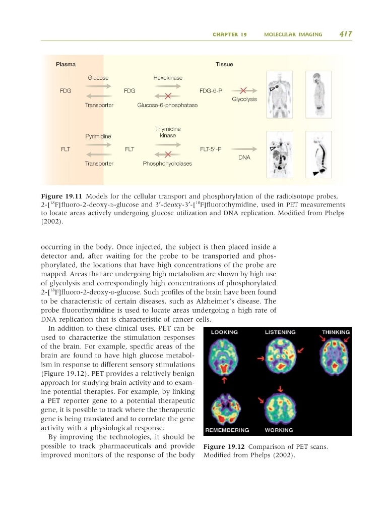
occurring in the body. Once injected, the subject is then placed inside a
detector and, after waiting for the probe to be transported and phosphorylated, the locations that have high concentrations of the probe are
mapped. Areas that are undergoing high metabolism are shown by high use
of glycolysis and correspondingly high concentrations of phosphorylated
2-[
18
F]fluoro-2-deoxy-D-glucose. Such profiles of the brain have been found
to be characteristic of certain diseases, such as Alzheimer’s disease. The
probe fluorothymidine is used to locate areas undergoing a high rate of
DNA replication that is characteristic of cancer cells.
In addition to these clinical uses, PET can be
used to characterize the stimulation responses
of the brain. For example, specific areas of the
brain are found to have high glucose metabolism in response to different sensory stimulations
(Figure 19.12). PET provides a relatively benign
approach for studying brain activity and to examine potential therapies. For example, by linking
a PET reporter gene to a potential therapeutic
gene, it is possible to track where the therapeutic
gene is being translated and to correlate the gene
activity with a physiological response.
By improving the technologies, it should be
possible to track pharmaceuticals and provide
improved monitors of the response of the body
Figure 19.11 Models for the cellular transport and phosphorylation of the radioisotope probes,
2-[
18 F]fluoro-2-deoxy-D-glucose and 3′-deoxy-3′-[
18 F]fluorothymidine, used in PET measurements
to locate areas actively undergoing glucose utilization and DNA replication. Modified from Phelps
(2002).
CHAPTER 19
MOLECULAR IMAGING
417
FDG
FLT
FDG
FLT
FDG-6-P
FLT-5Ј-P
Glucose
Transporter
Pyrimidine
Transporter
Hexokinase
Glucose-6-phosphatase
Thymidine
kinase
Phosphohydrolases
Glycolysis
DNA
Figure 19.12 Comparison of PET scans.
Modified from Phelps (2002).
9781405124362_4_019.qxd 4/29/08 14:08 Page 417
detector and, after waiting for the probe to be transported and phosphorylated, the locations that have high concentrations of the probe are
mapped. Areas that are undergoing high metabolism are shown by high use
of glycolysis and correspondingly high concentrations of phosphorylated
2-[
18
F]fluoro-2-deoxy-D-glucose. Such profiles of the brain have been found
to be characteristic of certain diseases, such as Alzheimer’s disease. The
probe fluorothymidine is used to locate areas undergoing a high rate of
DNA replication that is characteristic of cancer cells.
In addition to these clinical uses, PET can be
used to characterize the stimulation responses
of the brain. For example, specific areas of the
brain are found to have high glucose metabolism in response to different sensory stimulations
(Figure 19.12). PET provides a relatively benign
approach for studying brain activity and to examine potential therapies. For example, by linking
a PET reporter gene to a potential therapeutic
gene, it is possible to track where the therapeutic
gene is being translated and to correlate the gene
activity with a physiological response.
By improving the technologies, it should be
possible to track pharmaceuticals and provide
improved monitors of the response of the body
Figure 19.11 Models for the cellular transport and phosphorylation of the radioisotope probes,
2-[
18 F]fluoro-2-deoxy-D-glucose and 3′-deoxy-3′-[
18 F]fluorothymidine, used in PET measurements
to locate areas actively undergoing glucose utilization and DNA replication. Modified from Phelps
(2002).
CHAPTER 19
MOLECULAR IMAGING
417
FDG
FLT
FDG
FLT
FDG-6-P
FLT-5Ј-P
Glucose
Transporter
Pyrimidine
Transporter
Hexokinase
Glucose-6-phosphatase
Thymidine
kinase
Phosphohydrolases
Glycolysis
DNA
Figure 19.12 Comparison of PET scans.
Modified from Phelps (2002).
9781405124362_4_019.qxd 4/29/08 14:08 Page 417