

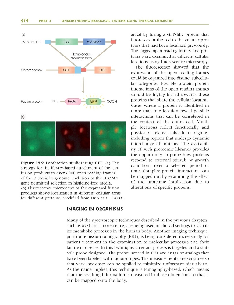
aided by fusing a GFP-like protein that
fluoresces in the red to the cellular proteins that had been localized previously.
The tagged open reading frames and proteins were examined at different cellular
locations using fluorescence microscopy.
The fluorescence showed that the
expression of the open reading frames
could be organized into distinct subcellular categories. Possible protein–protein
interactions of the open reading frames
should be highly biased towards those
proteins that share the cellular location.
Cases where a protein is identified in
more than one location reveal possible
interactions that can be considered in
the context of the entire cell. Multiple locations reflect functionally and
physically related subcellular regions,
including regions that undergo dynamic
interchange of proteins. The availability of such proteomic libraries provides
the opportunity to probe how proteins
respond to external stimuli or growth
conditions over a selected period of
time. Complex protein interactions can
be mapped out by examining the effect
of the proteome localization due to
alterations of specific proteins.
IMAGING IN ORGANISMS
Many of the spectroscopic techniques described in the previous chapters,
such as MRI and fluorescence, are being used in clinical settings to visualize metabolic processes in the human body. Another imaging technique,
positron emission tomography (PET), is being considered increasingly for
patient treatment in the examination of molecular processes and their
failure in disease. In this technique, a certain process is targeted and a suitable probe designed. The probes sensed in PET are drugs or analogs that
have been labeled with radioisotopes. The measurements are sensitive so
that very low doses can be applied to minimize unforeseen side effects.
As the name implies, this technique is tomography-based, which means
that the resulting information is measured in three dimensions so that it
can be mapped onto the body.
414
PART 3
UNDERSTANDING BIOLOGICAL SYSTEMS USING PHYSICAL CHEMISTRY
PCR product
(a)
Chromosome
Homologous
recombination
Fusion protein
Protein
COOH
NH 2
ORF
ORF
GFP
HIS3MX6
GFP
Figure 19.9 Localization studies using GFP. (a) The
strategy for the library-based attachment of the GFP
fusion products to over 6000 open reading frames
of the S. cerevisiae genome. Inclusion of the His3MX
gene permitted selection in histidine-free media.
(b) Fluorescence microscopy of the expressed fusion
products shows localization in different cellular areas
for different proteins. Modified from Huh et al. (2003).
9781405124362_4_019.qxd 4/29/08 14:07 Page 414
fluoresces in the red to the cellular proteins that had been localized previously.
The tagged open reading frames and proteins were examined at different cellular
locations using fluorescence microscopy.
The fluorescence showed that the
expression of the open reading frames
could be organized into distinct subcellular categories. Possible protein–protein
interactions of the open reading frames
should be highly biased towards those
proteins that share the cellular location.
Cases where a protein is identified in
more than one location reveal possible
interactions that can be considered in
the context of the entire cell. Multiple locations reflect functionally and
physically related subcellular regions,
including regions that undergo dynamic
interchange of proteins. The availability of such proteomic libraries provides
the opportunity to probe how proteins
respond to external stimuli or growth
conditions over a selected period of
time. Complex protein interactions can
be mapped out by examining the effect
of the proteome localization due to
alterations of specific proteins.
IMAGING IN ORGANISMS
Many of the spectroscopic techniques described in the previous chapters,
such as MRI and fluorescence, are being used in clinical settings to visualize metabolic processes in the human body. Another imaging technique,
positron emission tomography (PET), is being considered increasingly for
patient treatment in the examination of molecular processes and their
failure in disease. In this technique, a certain process is targeted and a suitable probe designed. The probes sensed in PET are drugs or analogs that
have been labeled with radioisotopes. The measurements are sensitive so
that very low doses can be applied to minimize unforeseen side effects.
As the name implies, this technique is tomography-based, which means
that the resulting information is measured in three dimensions so that it
can be mapped onto the body.
414
PART 3
UNDERSTANDING BIOLOGICAL SYSTEMS USING PHYSICAL CHEMISTRY
PCR product
(a)
Chromosome
Homologous
recombination
Fusion protein
Protein
COOH
NH 2
ORF
ORF
GFP
HIS3MX6
GFP
Figure 19.9 Localization studies using GFP. (a) The
strategy for the library-based attachment of the GFP
fusion products to over 6000 open reading frames
of the S. cerevisiae genome. Inclusion of the His3MX
gene permitted selection in histidine-free media.
(b) Fluorescence microscopy of the expressed fusion
products shows localization in different cellular areas
for different proteins. Modified from Huh et al. (2003).
9781405124362_4_019.qxd 4/29/08 14:07 Page 414