

CHAPTER 19
MOLECULAR IMAGING
409
A
B
0.2
0.4
0.6
0.8
1.0
(a)
0.0
300
350
400
450
500
550
Absorbance
Wavelength (nm)
0.2
0.4
0.6
0.8
1.0
(b)
0.0
5
6
7
8
9
Normalized fluorescence
pH
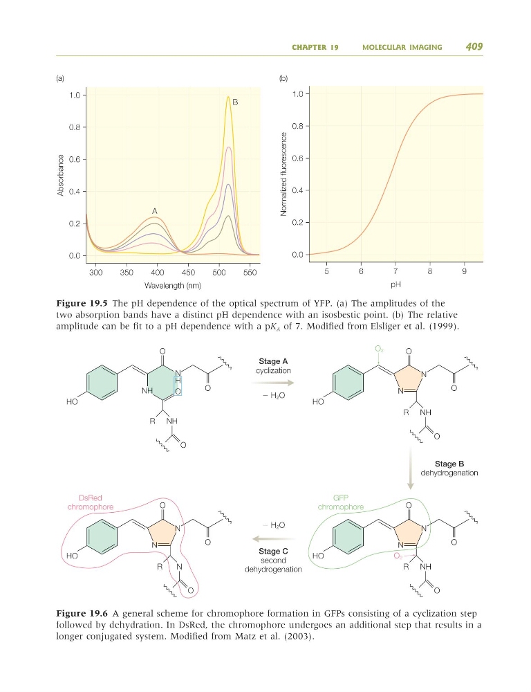
Figure 19.5 The pH dependence of the optical spectrum of YFP. (a) The amplitudes of the
two absorption bands have a distinct pH dependence with an isosbestic point. (b) The relative
amplitude can be fit to a pH dependence with a pK A of 7. Modified from Elsliger et al. (1999).
O
O
cyclization
Ϫ H 2O
Ϫ H2O
dehydrogenation
second
dehydrogenation
DsRed
chromophore
GFP
chromophore
HO
R
O
NH
O
O2
O
HO
N
N
R
O
NH
O
O
HO
N
N
R
O
N
O
O
HO
N
N
R
O 2
O
NH
N
NH
H
O
Figure 19.6 A general scheme for chromophore formation in GFPs consisting of a cyclization step
followed by dehydration. In DsRed, the chromophore undergoes an additional step that results in a
longer conjugated system. Modified from Matz et al. (2003).
9781405124362_4_019.qxd 4/29/08 14:07 Page 409
MOLECULAR IMAGING
409
A
B
0.2
0.4
0.6
0.8
1.0
(a)
0.0
300
350
400
450
500
550
Absorbance
Wavelength (nm)
0.2
0.4
0.6
0.8
1.0
(b)
0.0
5
6
7
8
9
Normalized fluorescence
pH
Figure 19.5 The pH dependence of the optical spectrum of YFP. (a) The amplitudes of the
two absorption bands have a distinct pH dependence with an isosbestic point. (b) The relative
amplitude can be fit to a pH dependence with a pK A of 7. Modified from Elsliger et al. (1999).
O
O
cyclization
Ϫ H 2O
Ϫ H2O
dehydrogenation
second
dehydrogenation
DsRed
chromophore
GFP
chromophore
HO
R
O
NH
O
O2
O
HO
N
N
R
O
NH
O
O
HO
N
N
R
O
N
O
O
HO
N
N
R
O 2
O
NH
N
NH
H
O
Figure 19.6 A general scheme for chromophore formation in GFPs consisting of a cyclization step
followed by dehydration. In DsRed, the chromophore undergoes an additional step that results in a
longer conjugated system. Modified from Matz et al. (2003).
9781405124362_4_019.qxd 4/29/08 14:07 Page 409