

which grows in briny waters such as the Dead
Sea. This bacterium has a distinctive purple
color due to the presence of the extremely high
concentrations of retinal found in bacteriorhodopsin. The bacteriorhodopsin is present,
not for a visual response such as phototaxis,
but rather to convert light energy into chemical
energy in the form of proton gradients across the
cytoplasmic membrane.
Both rhodopsin and bacteriorhodopsin are proteins with a simple polypeptide chain formed
by about 250 amino acid residues. Many of the
amino acid residues of these polypeptides are
conserved between rhodopsin and bacteriorhodopsin. As was found for rhodopsin, a retinal
binds through a Schiff base to a lysine, which is
residue 216 in bacteriorhodopsin. The photocycle of bacteriorhodopsin has similarities to
that of rhodopsin, with the presence of different spectral states that are generated on varying
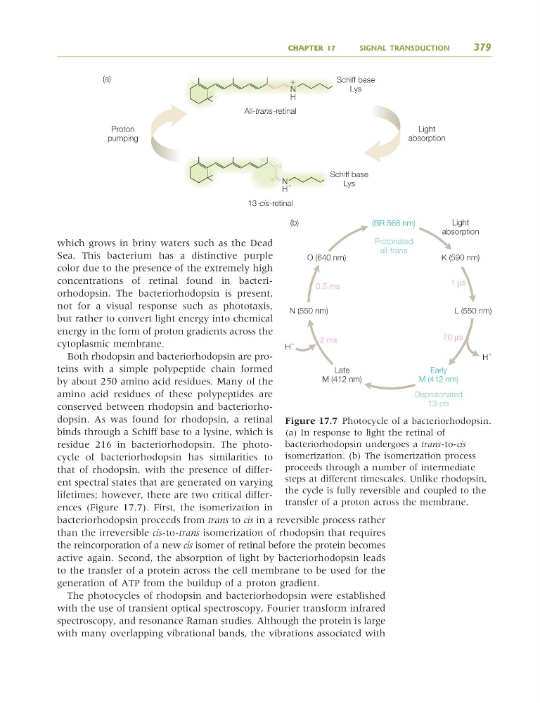
lifetimes; however, there are two critical differences (Figure 17.7). First, the isomerization in
bacteriorhodopsin proceeds from trans to cis in a reversible process rather
than the irreversible cis-to-trans isomerization of rhodopsin that requires
the reincorporation of a new cis isomer of retinal before the protein becomes
active again. Second, the absorption of light by bacteriorhodopsin leads
to the transfer of a protein across the cell membrane to be used for the
generation of ATP from the buildup of a proton gradient.
The photocycles of rhodopsin and bacteriorhodopsin were established
with the use of transient optical spectroscopy, Fourier transform infrared
spectroscopy, and resonance Raman studies. Although the protein is large
with many overlapping vibrational bands, the vibrations associated with
CHAPTER 17
SIGNAL TRANSDUCTION
379
All-trans-retinal
N
Schiff base
Lys
Schiff base
Lys
H
ϩ
Light
absorption
Proton
pumping
(a)
13-cis-retinal
N
H
ϩ
13
13
15
(BR 568 nm)
Protonated
all-trans
Deprotonated
13-cis
Light
absorption
K (590 nm)
L (550 nm)
Early
M (412 nm)
Late
M (412 nm)
N (550 nm)
O (640 nm)
(b)
0.5 ms
1 μs
70 μs
2 ms
H
ϩ
H
ϩ
Figure 17.7 Photocycle of a bacteriorhodopsin.
(a) In response to light the retinal of
bacteriorhodopsin undergoes a trans-to-cis
isomerization. (b) The isomerization process
proceeds through a number of intermediate
steps at different timescales. Unlike rhodopsin,
the cycle is fully reversible and coupled to the
transfer of a proton across the membrane.
9781405124362_4_017.qxd 4/29/08 10:46 Page 379
Sea. This bacterium has a distinctive purple
color due to the presence of the extremely high
concentrations of retinal found in bacteriorhodopsin. The bacteriorhodopsin is present,
not for a visual response such as phototaxis,
but rather to convert light energy into chemical
energy in the form of proton gradients across the
cytoplasmic membrane.
Both rhodopsin and bacteriorhodopsin are proteins with a simple polypeptide chain formed
by about 250 amino acid residues. Many of the
amino acid residues of these polypeptides are
conserved between rhodopsin and bacteriorhodopsin. As was found for rhodopsin, a retinal
binds through a Schiff base to a lysine, which is
residue 216 in bacteriorhodopsin. The photocycle of bacteriorhodopsin has similarities to
that of rhodopsin, with the presence of different spectral states that are generated on varying
lifetimes; however, there are two critical differences (Figure 17.7). First, the isomerization in
bacteriorhodopsin proceeds from trans to cis in a reversible process rather
than the irreversible cis-to-trans isomerization of rhodopsin that requires
the reincorporation of a new cis isomer of retinal before the protein becomes
active again. Second, the absorption of light by bacteriorhodopsin leads
to the transfer of a protein across the cell membrane to be used for the
generation of ATP from the buildup of a proton gradient.
The photocycles of rhodopsin and bacteriorhodopsin were established
with the use of transient optical spectroscopy, Fourier transform infrared
spectroscopy, and resonance Raman studies. Although the protein is large
with many overlapping vibrational bands, the vibrations associated with
CHAPTER 17
SIGNAL TRANSDUCTION
379
All-trans-retinal
N
Schiff base
Lys
Schiff base
Lys
H
ϩ
Light
absorption
Proton
pumping
(a)
13-cis-retinal
N
H
ϩ
13
13
15
(BR 568 nm)
Protonated
all-trans
Deprotonated
13-cis
Light
absorption
K (590 nm)
L (550 nm)
Early
M (412 nm)
Late
M (412 nm)
N (550 nm)
O (640 nm)
(b)
0.5 ms
1 μs
70 μs
2 ms
H
ϩ
H
ϩ
Figure 17.7 Photocycle of a bacteriorhodopsin.
(a) In response to light the retinal of
bacteriorhodopsin undergoes a trans-to-cis
isomerization. (b) The isomerization process
proceeds through a number of intermediate
steps at different timescales. Unlike rhodopsin,
the cycle is fully reversible and coupled to the
transfer of a proton across the membrane.
9781405124362_4_017.qxd 4/29/08 10:46 Page 379