

biological signal transduction as it involves the
absorption of light by a pigment buried inside of
a protein called rhodopsin rather than the binding of a signal molecule to a protein receptor, as
found in other signal-transduction processes. After
recognition, there is a signal conversion, which
requires a structural change of the protein and
an associated molecule called retinal in response
to light absorption. Once the signal has activated
rhodopsin, it is amplified by many orders of magnitude, allowing the signal to be processed into a
change in membrane potential and consequently
a signal to the brain.
On a biological level, these four steps are
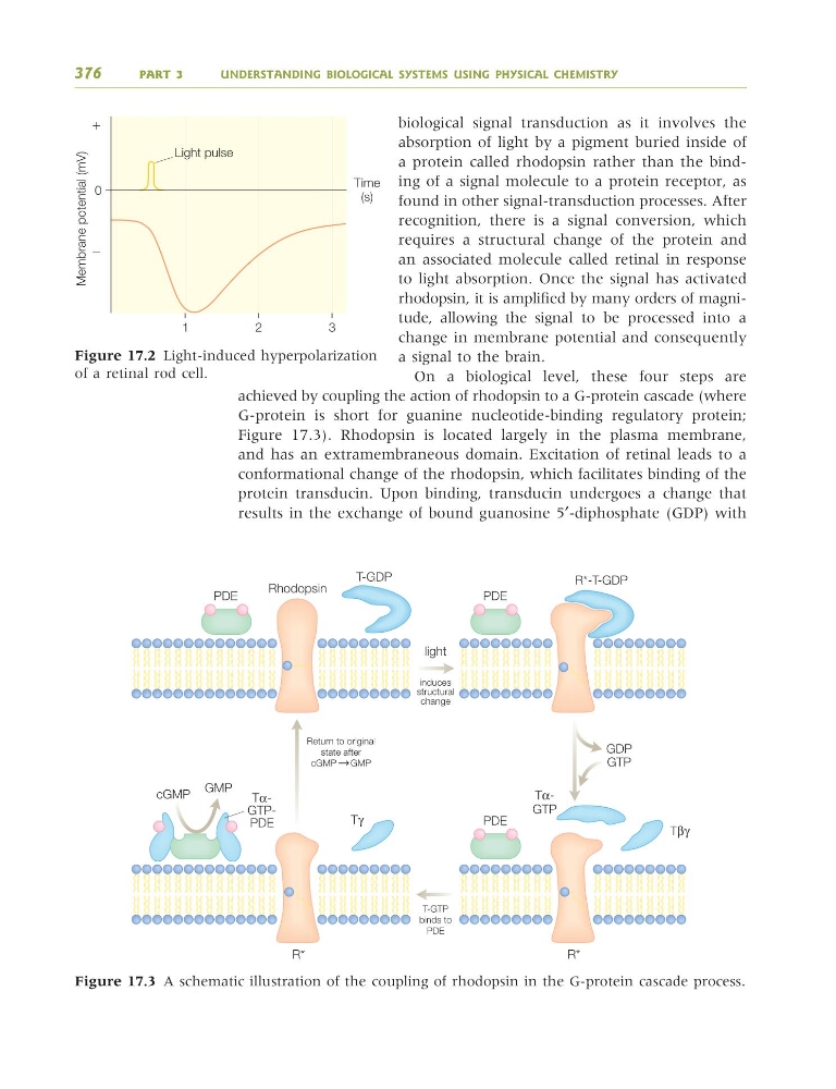
achieved by coupling the action of rhodopsin to a G-protein cascade (where
G-protein is short for guanine nucleotide-binding regulatory protein;
Figure 17.3). Rhodopsin is located largely in the plasma membrane,
and has an extramembraneous domain. Excitation of retinal leads to a
conformational change of the rhodopsin, which facilitates binding of the
protein transducin. Upon binding, transducin undergoes a change that
results in the exchange of bound guanosine 5′-diphosphate (GDP) with
376
PART 3
UNDERSTANDING BIOLOGICAL SYSTEMS USING PHYSICAL CHEMISTRY
Time
(s)
Light pulse
0
ϩ
Ϫ
1
2
3
Membrane potential (mV)
Figure 17.2 Light-induced hyperpolarization
of a retinal rod cell.
PDE
PDE
PDE
cGMP
GMP
GDP
GTP
Rhodopsin
T-GDP
R*-T-GDP
R*
R*
Tβγ
Tγ
TαGTP
light
induces
structural
change
Return to original
state after
cGMP GMP
T-GTP
binds to
PDE
GTPPDE
Figure 17.3 A schematic illustration of the coupling of rhodopsin in the G-protein cascade process.
9781405124362_4_017.qxd 4/29/08 10:46 Page 376
absorption of light by a pigment buried inside of
a protein called rhodopsin rather than the binding of a signal molecule to a protein receptor, as
found in other signal-transduction processes. After
recognition, there is a signal conversion, which
requires a structural change of the protein and
an associated molecule called retinal in response
to light absorption. Once the signal has activated
rhodopsin, it is amplified by many orders of magnitude, allowing the signal to be processed into a
change in membrane potential and consequently
a signal to the brain.
On a biological level, these four steps are
achieved by coupling the action of rhodopsin to a G-protein cascade (where
G-protein is short for guanine nucleotide-binding regulatory protein;
Figure 17.3). Rhodopsin is located largely in the plasma membrane,
and has an extramembraneous domain. Excitation of retinal leads to a
conformational change of the rhodopsin, which facilitates binding of the
protein transducin. Upon binding, transducin undergoes a change that
results in the exchange of bound guanosine 5′-diphosphate (GDP) with
376
PART 3
UNDERSTANDING BIOLOGICAL SYSTEMS USING PHYSICAL CHEMISTRY
Time
(s)
Light pulse
0
ϩ
Ϫ
1
2
3
Membrane potential (mV)
Figure 17.2 Light-induced hyperpolarization
of a retinal rod cell.
PDE
PDE
PDE
cGMP
GMP
GDP
GTP
Rhodopsin
T-GDP
R*-T-GDP
R*
R*
Tβγ
Tγ
TαGTP
light
induces
structural
change
Return to original
state after
cGMP GMP
T-GTP
binds to
PDE
GTPPDE
Figure 17.3 A schematic illustration of the coupling of rhodopsin in the G-protein cascade process.
9781405124362_4_017.qxd 4/29/08 10:46 Page 376