

structures if the structures are not too large. Although NMR techniques
are being developed to overcome this limitation, the upper boundary
for normal NMR spectroscopy is a molecular mass of about 50,000 Da.
Therefore, an active area of research is the development of novel techniques that can be used for larger structures (Riek et al. 2000; Hakumaki
& Brindle 2003; Tugarinov et al. 2004; Vaynberg & Qin 2006). In conventional two-dimensional NMR, the spectra of large proteins typically
CHAPTER 16
MAGNETIC RESONANCE
353
(a)
3
2
1
4
4
CαH
CβH3
3
2
1
ppm
ppm
Alanine
1.3
4.35
CβH3
H
Cα
COO
Ϫ
ND2
(b)
3
2
1
4
4
3
2
1
ppm
ppm
CδH3
CδH3
CδH3
L-Leucine
H
Cγ
CβH2
H
Cα
CαH
CβH2
CγH
COO
Ϫ
ND2
(c)
3
2
1
4
4
3
2
1
ppm
ppm
H
Threonine
DO Cβ
CγH3
CγH3
H
Cα
CβH
CαH
COO
Ϫ
ND2
(d)
3
2
1
4
4
CαH
CβH
CγH3
CγH3
3
2
1
ppm
ppm
Valine
H
Cα
COO
Ϫ
ND2
CγH3
H
Cβ
CγH3
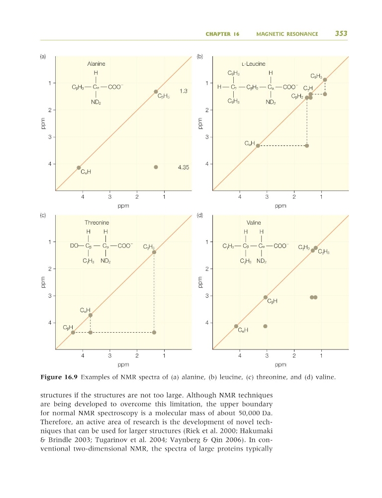
Figure 16.9 Examples of NMR spectra of (a) alanine, (b) leucine, (c) threonine, and (d) valine.
9781405124362_4_016.qxd 4/29/08 10:45 Page 353
are being developed to overcome this limitation, the upper boundary
for normal NMR spectroscopy is a molecular mass of about 50,000 Da.
Therefore, an active area of research is the development of novel techniques that can be used for larger structures (Riek et al. 2000; Hakumaki
& Brindle 2003; Tugarinov et al. 2004; Vaynberg & Qin 2006). In conventional two-dimensional NMR, the spectra of large proteins typically
CHAPTER 16
MAGNETIC RESONANCE
353
(a)
3
2
1
4
4
CαH
CβH3
3
2
1
ppm
ppm
Alanine
1.3
4.35
CβH3
H
Cα
COO
Ϫ
ND2
(b)
3
2
1
4
4
3
2
1
ppm
ppm
CδH3
CδH3
CδH3
L-Leucine
H
Cγ
CβH2
H
Cα
CαH
CβH2
CγH
COO
Ϫ
ND2
(c)
3
2
1
4
4
3
2
1
ppm
ppm
H
Threonine
DO Cβ
CγH3
CγH3
H
Cα
CβH
CαH
COO
Ϫ
ND2
(d)
3
2
1
4
4
CαH
CβH
CγH3
CγH3
3
2
1
ppm
ppm
Valine
H
Cα
COO
Ϫ
ND2
CγH3
H
Cβ
CγH3
Figure 16.9 Examples of NMR spectra of (a) alanine, (b) leucine, (c) threonine, and (d) valine.
9781405124362_4_016.qxd 4/29/08 10:45 Page 353