

The variable part of the chain is the identity of the base at each position,
or the sequence.
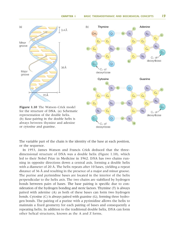
In 1953, James Watson and Francis Crick deduced that the threedimensional structure of DNA was a double helix (Figure 1.10), which
led to their Nobel Prize in Medicine in 1962. DNA has two chains running in opposite directions down a central axis, forming a double helix
with a diameter of 20 Å. The helix repeats after 10 bases, yielding a repeat
distance of 36 Å and resulting in the presence of a major and minor groove.
The purine and pyrimidine bases are located in the interior of the helix
perpendicular to the helix axis. The two chains are stabilized by hydrogen
bonds between pairs of bases. The base pairing is specific due to consideration of the hydrogen bonding and steric factors. Thymine (T) is always
paired with adenine (A) as both of these bases can form two hydrogen
bonds. Cytosine (C) is always paired with guanine (G), forming three hydrogen bonds. The pairing of a purine with a pyrimidine allows the helix to
maintain a fixed geometry for each pairing of bases and consequently a
repeating helix. In addition to the traditional double helix, DNA can form
other helical structures, known as the A and Z forms.
Figure 1.10 The Watson–Crick model
for the structure of DNA. (a) Schematic
representation of the double helix.
(b) Base-pairing in the double helix is
always between thymine and adenine
or cytosine and guanine.
CHAPTER 1
BASIC THERMODYNAMIC AND BIOCHEMICAL CONCEPTS
19
3.4 Å
36 Å
20 Å
Major
groove
(a)
Minor
groove
(b)
Thymine
Adenine
C1Ј of
deoxyribose
C 1Ј of
deoxyribose
CH3
O
C6
H
H
C5
H
H
H
H
N
N1
C6
C2
C4
N1
C2
N3
N7
N9
C5
C8
N3
C1Ј
C4
C1Ј
O
Cytosine
Guanine
N
C6
H
H
C5
H
N
H
H
H
H
H
O
N1
C6
C2
C4
N1
C2
N3
N7
N9
C5
C8
N3
C1Ј
C4
C1Ј
O
C1Ј of
deoxyribose
C 1Ј of
deoxyribose
9781405124362_4_001.qxd 4/29/08 9:07 Page 19
or the sequence.
In 1953, James Watson and Francis Crick deduced that the threedimensional structure of DNA was a double helix (Figure 1.10), which
led to their Nobel Prize in Medicine in 1962. DNA has two chains running in opposite directions down a central axis, forming a double helix
with a diameter of 20 Å. The helix repeats after 10 bases, yielding a repeat
distance of 36 Å and resulting in the presence of a major and minor groove.
The purine and pyrimidine bases are located in the interior of the helix
perpendicular to the helix axis. The two chains are stabilized by hydrogen
bonds between pairs of bases. The base pairing is specific due to consideration of the hydrogen bonding and steric factors. Thymine (T) is always
paired with adenine (A) as both of these bases can form two hydrogen
bonds. Cytosine (C) is always paired with guanine (G), forming three hydrogen bonds. The pairing of a purine with a pyrimidine allows the helix to
maintain a fixed geometry for each pairing of bases and consequently a
repeating helix. In addition to the traditional double helix, DNA can form
other helical structures, known as the A and Z forms.
Figure 1.10 The Watson–Crick model
for the structure of DNA. (a) Schematic
representation of the double helix.
(b) Base-pairing in the double helix is
always between thymine and adenine
or cytosine and guanine.
CHAPTER 1
BASIC THERMODYNAMIC AND BIOCHEMICAL CONCEPTS
19
3.4 Å
36 Å
20 Å
Major
groove
(a)
Minor
groove
(b)
Thymine
Adenine
C1Ј of
deoxyribose
C 1Ј of
deoxyribose
CH3
O
C6
H
H
C5
H
H
H
H
N
N1
C6
C2
C4
N1
C2
N3
N7
N9
C5
C8
N3
C1Ј
C4
C1Ј
O
Cytosine
Guanine
N
C6
H
H
C5
H
N
H
H
H
H
H
O
N1
C6
C2
C4
N1
C2
N3
N7
N9
C5
C8
N3
C1Ј
C4
C1Ј
O
C1Ј of
deoxyribose
C 1Ј of
deoxyribose
9781405124362_4_001.qxd 4/29/08 9:07 Page 19