

of very real importance as such aggregates are
linked to many diseases that cannot be treated,
including Alzheimer’s disease, Creutzfeldt–
Jakob disease, and bovine spongiform encephalopathy (so-called mad-cow disease). The
involvement of misfolded proteins giving rise
to these diseases was initially met with skepticism, but the efforts of Stanley Pruisner in the
1980s (Prusiner 1987) have led to a general
acceptance as recognized by a Nobel Prize in
Medicine in 1997.
Whereas these proteins, which are termed
prions, for proteinaceous infectious particles,
usually have a globular shape, they can also
adopt a structure that leads to formation of
amyloid-like fibrils (Zahn et al. 2000; Dobson
2003; Masison 2004; May et al. 2004; Dyson
& Wright 2005; Krishnan & Lindquist 2005;
Nelson et al. 2005). Proteins in amyloid fibrils
are folded to form continuous arrays of β sheets.
A large portion of a prion is folded in a compact globular arrangement with α helices and
β sheets, with a sizable portion of the protein
missing due to disorder, including most of
the first 100 amino acid residues (Zahn et al.
2000; Figure 8.5).
The determination of the arrangement of
prions in amyloid fibrils has been hampered
by the limited order of fibrils isolated from
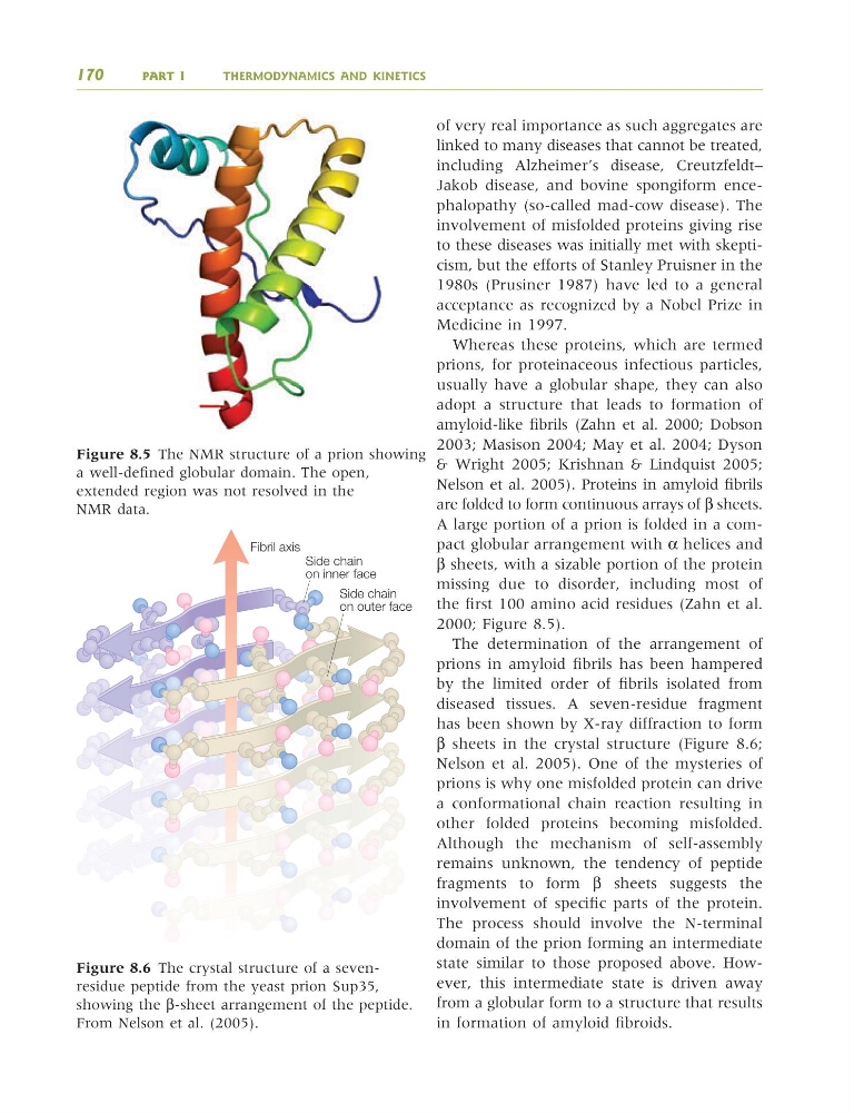
diseased tissues. A seven-residue fragment
has been shown by X-ray diffraction to form
β sheets in the crystal structure (Figure 8.6;
Nelson et al. 2005). One of the mysteries of
prions is why one misfolded protein can drive
a conformational chain reaction resulting in
other folded proteins becoming misfolded.
Although the mechanism of self-assembly
remains unknown, the tendency of peptide
fragments to form β sheets suggests the
involvement of specific parts of the protein.
The process should involve the N-terminal
domain of the prion forming an intermediate
state similar to those proposed above. However, this intermediate state is driven away
from a globular form to a structure that results
in formation of amyloid fibroids.
170
PART I
THERMODYNAMICS AND KINETICS
Side chain
on inner face
Side chain
on outer face
Fibril axis
Figure 8.6 The crystal structure of a sevenresidue peptide from the yeast prion Sup35,
showing the β-sheet arrangement of the peptide.
From Nelson et al. (2005).
Figure 8.5 The NMR structure of a prion showing
a well-defined globular domain. The open,
extended region was not resolved in the
NMR data.
9781405124362_4_008.qxd 4/29/08 9:09 Page 170
linked to many diseases that cannot be treated,
including Alzheimer’s disease, Creutzfeldt–
Jakob disease, and bovine spongiform encephalopathy (so-called mad-cow disease). The
involvement of misfolded proteins giving rise
to these diseases was initially met with skepticism, but the efforts of Stanley Pruisner in the
1980s (Prusiner 1987) have led to a general
acceptance as recognized by a Nobel Prize in
Medicine in 1997.
Whereas these proteins, which are termed
prions, for proteinaceous infectious particles,
usually have a globular shape, they can also
adopt a structure that leads to formation of
amyloid-like fibrils (Zahn et al. 2000; Dobson
2003; Masison 2004; May et al. 2004; Dyson
& Wright 2005; Krishnan & Lindquist 2005;
Nelson et al. 2005). Proteins in amyloid fibrils
are folded to form continuous arrays of β sheets.
A large portion of a prion is folded in a compact globular arrangement with α helices and
β sheets, with a sizable portion of the protein
missing due to disorder, including most of
the first 100 amino acid residues (Zahn et al.
2000; Figure 8.5).
The determination of the arrangement of
prions in amyloid fibrils has been hampered
by the limited order of fibrils isolated from
diseased tissues. A seven-residue fragment
has been shown by X-ray diffraction to form
β sheets in the crystal structure (Figure 8.6;
Nelson et al. 2005). One of the mysteries of
prions is why one misfolded protein can drive
a conformational chain reaction resulting in
other folded proteins becoming misfolded.
Although the mechanism of self-assembly
remains unknown, the tendency of peptide
fragments to form β sheets suggests the
involvement of specific parts of the protein.
The process should involve the N-terminal
domain of the prion forming an intermediate
state similar to those proposed above. However, this intermediate state is driven away
from a globular form to a structure that results
in formation of amyloid fibroids.
170
PART I
THERMODYNAMICS AND KINETICS
Side chain
on inner face
Side chain
on outer face
Fibril axis
Figure 8.6 The crystal structure of a sevenresidue peptide from the yeast prion Sup35,
showing the β-sheet arrangement of the peptide.
From Nelson et al. (2005).
Figure 8.5 The NMR structure of a prion showing
a well-defined globular domain. The open,
extended region was not resolved in the
NMR data.
9781405124362_4_008.qxd 4/29/08 9:09 Page 170