

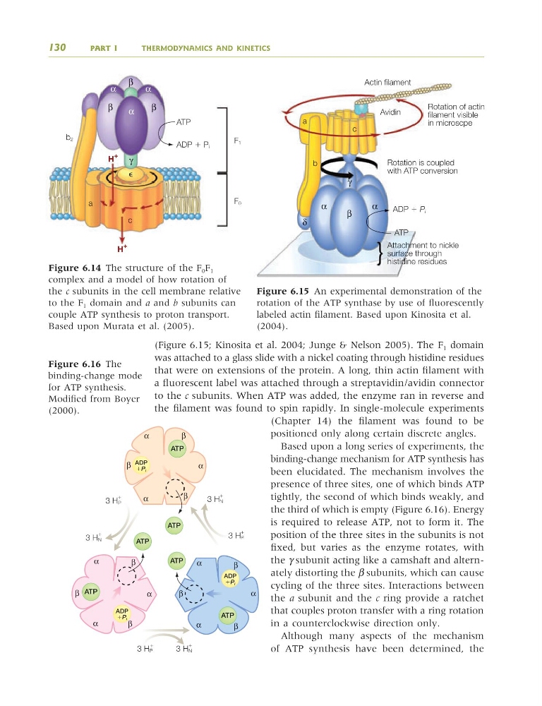
(Figure 6.15; Kinosita et al. 2004; Junge & Nelson 2005). The F 1 domain
was attached to a glass slide with a nickel coating through histidine residues
that were on extensions of the protein. A long, thin actin filament with
a fluorescent label was attached through a streptavidin/avidin connector
to the c subunits. When ATP was added, the enzyme ran in reverse and
the filament was found to spin rapidly. In single-molecule experiments
(Chapter 14) the filament was found to be
positioned only along certain discrete angles.
Based upon a long series of experiments, the
binding-change mechanism for ATP synthesis has
been elucidated. The mechanism involves the
presence of three sites, one of which binds ATP
tightly, the second of which binds weakly, and
the third of which is empty (Figure 6.16). Energy
is required to release ATP, not to form it. The
position of the three sites in the subunits is not
fixed, but varies as the enzyme rotates, with
the γ subunit acting like a camshaft and alternately distorting the β subunits, which can cause
cycling of the three sites. Interactions between
the a subunit and the c ring provide a ratchet
that couples proton transfer with a ring rotation
in a counterclockwise direction only.
Although many aspects of the mechanism
of ATP synthesis have been determined, the
130
PART I
THERMODYNAMICS AND KINETICS
γ
δ
α
β
α
Attachment to nickle
surface through
histidine residues
Rotation is coupled
with ATP conversion
Rotation of actin
filament visible
in microscpe
Avidin
Actin filament
a
c
b
ATP
ADP ϩ
ATP
ATP
ATP
ATP
ATP
ATP
ADP
ϩPi
ADP
ϩPi
ADP
ϩPi
ϩ
ϩ
ϩ
3 HN
3 HP
α
α
α
α
α
α
α
β
β
β
β
β
β
β
β
β
α
α
3 HN
ϩ
3 HN
ϩ
3 HP
3 HP
ϩ
Figure 6.15 An experimental demonstration of the
rotation of the ATP synthase by use of fluorescently
labeled actin filament. Based upon Kinosita et al.
(2004).
Figure 6.16 The
binding-change mode
for ATP synthesis.
Modified from Boyer
(2000).
β
β
β
γ
⑀
α
α
α
F1
ATP
b2
a
c
H
ϩ ϩ
H
ϩ
ADP ϩ Pi
F0
Figure 6.14 The structure of the F 0 F 1
complex and a model of how rotation of
the c subunits in the cell membrane relative
to the F 1 domain and a and b subunits can
couple ATP synthesis to proton transport.
Based upon Murata et al. (2005).
9781405124362_4_006.qxd 4/29/08 14:07 Page 130
was attached to a glass slide with a nickel coating through histidine residues
that were on extensions of the protein. A long, thin actin filament with
a fluorescent label was attached through a streptavidin/avidin connector
to the c subunits. When ATP was added, the enzyme ran in reverse and
the filament was found to spin rapidly. In single-molecule experiments
(Chapter 14) the filament was found to be
positioned only along certain discrete angles.
Based upon a long series of experiments, the
binding-change mechanism for ATP synthesis has
been elucidated. The mechanism involves the
presence of three sites, one of which binds ATP
tightly, the second of which binds weakly, and
the third of which is empty (Figure 6.16). Energy
is required to release ATP, not to form it. The
position of the three sites in the subunits is not
fixed, but varies as the enzyme rotates, with
the γ subunit acting like a camshaft and alternately distorting the β subunits, which can cause
cycling of the three sites. Interactions between
the a subunit and the c ring provide a ratchet
that couples proton transfer with a ring rotation
in a counterclockwise direction only.
Although many aspects of the mechanism
of ATP synthesis have been determined, the
130
PART I
THERMODYNAMICS AND KINETICS
γ
δ
α
β
α
Attachment to nickle
surface through
histidine residues
Rotation is coupled
with ATP conversion
Rotation of actin
filament visible
in microscpe
Avidin
Actin filament
a
c
b
ATP
ADP ϩ
ATP
ATP
ATP
ATP
ATP
ATP
ADP
ϩPi
ADP
ϩPi
ADP
ϩPi
ϩ
ϩ
ϩ
3 HN
3 HP
α
α
α
α
α
α
α
β
β
β
β
β
β
β
β
β
α
α
3 HN
ϩ
3 HN
ϩ
3 HP
3 HP
ϩ
Figure 6.15 An experimental demonstration of the
rotation of the ATP synthase by use of fluorescently
labeled actin filament. Based upon Kinosita et al.
(2004).
Figure 6.16 The
binding-change mode
for ATP synthesis.
Modified from Boyer
(2000).
β
β
β
γ
⑀
α
α
α
F1
ATP
b2
a
c
H
ϩ ϩ
H
ϩ
ADP ϩ Pi
F0
Figure 6.14 The structure of the F 0 F 1
complex and a model of how rotation of
the c subunits in the cell membrane relative
to the F 1 domain and a and b subunits can
couple ATP synthesis to proton transport.
Based upon Murata et al. (2005).
9781405124362_4_006.qxd 4/29/08 14:07 Page 130