

360
Our work in this area was stimulated by observations made during surveys of the distribution
of Synechococcus, in which we sometimes noticed two populations of cells, having relatively
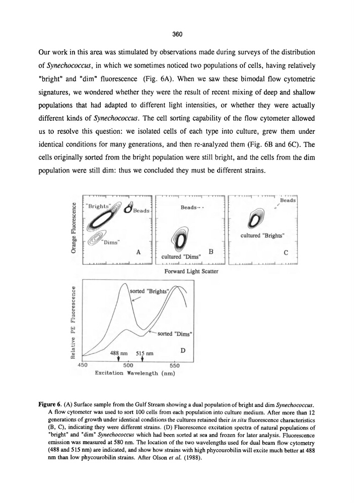
"bright" and "dim" fluorescence (Fig. 6A). When we saw these bimodal flow cytometric
signatures, we wondered whether they were the result of recent mixing of deep and shallow
populations that had adapted to different light intensities, or whether they were actually
different kinds of Synechococcus. The cell sorting capability of the flow cytometer allowed
us to resolve this question: we isolated cells of each type into culture, grew them under
identical conditions for many generations, and then re-analyzed them (Fig. 6B and 6C). The
cells originally sorted from the bright population were still bright, and the cells from the dim
population were still dim: thus we concluded they must be different strains.
III
U
e
III
u
OJ
III
. .
o
:l
r;:
-.:I
n..
dBead S
Beads- .
A
cultured "Dims"
8
L-...L.,. ,,,,I
!
f
Forward Light Scatter
550
Exci tation Wavelength (nm)
D
""I i" lIn
Beads
cultured "Brights"
c
",".1
. . 1".,,1
Figure 6. (A) Surface sample from the Gulf Stream showing a dual population of bright and dim Synechococcus.
A flow cytometer was used to sort 100 cells from each population into culture medium. After more than 12
generations of growth under identical conditions the cultures retained their ill situ fluorescence characteristics
(B, C), indicating they were different strains. (D) Fluorescence excitation spectra of natural popUlations of
"bright" and "dim" SYllechococcus which had been sorted at sea and frozen for later analysis. Fluorescence
emission was measured at 580 nm. The location of the two wavelengths used for dual beam flow cytometry
(488 and 515 nm) are indicated, and show how strains with high phycourobilin will excite much better at 488
nm than low phycourobilin strains. After Olson et al. (1988).
Our work in this area was stimulated by observations made during surveys of the distribution
of Synechococcus, in which we sometimes noticed two populations of cells, having relatively
"bright" and "dim" fluorescence (Fig. 6A). When we saw these bimodal flow cytometric
signatures, we wondered whether they were the result of recent mixing of deep and shallow
populations that had adapted to different light intensities, or whether they were actually
different kinds of Synechococcus. The cell sorting capability of the flow cytometer allowed
us to resolve this question: we isolated cells of each type into culture, grew them under
identical conditions for many generations, and then re-analyzed them (Fig. 6B and 6C). The
cells originally sorted from the bright population were still bright, and the cells from the dim
population were still dim: thus we concluded they must be different strains.
III
U
e
III
u
OJ
III
. .
o
:l
r;:
-.:I
n..
dBead S
Beads- .
A
cultured "Dims"
8
L-...L.,. ,,,,I
!
f
Forward Light Scatter
550
Exci tation Wavelength (nm)
D
""I i" lIn
Beads
cultured "Brights"
c
",".1
. . 1".,,1
Figure 6. (A) Surface sample from the Gulf Stream showing a dual population of bright and dim Synechococcus.
A flow cytometer was used to sort 100 cells from each population into culture medium. After more than 12
generations of growth under identical conditions the cultures retained their ill situ fluorescence characteristics
(B, C), indicating they were different strains. (D) Fluorescence excitation spectra of natural popUlations of
"bright" and "dim" SYllechococcus which had been sorted at sea and frozen for later analysis. Fluorescence
emission was measured at 580 nm. The location of the two wavelengths used for dual beam flow cytometry
(488 and 515 nm) are indicated, and show how strains with high phycourobilin will excite much better at 488
nm than low phycourobilin strains. After Olson et al. (1988).