

12
used to focus the excitation light. Hence, excitation and fluorescence collection are
automatically confocal with a common optical axis. This feature makes such instruments
inherently much more rugged than laser-based ones. It also greatly simplifies the alignment,
which is reduced to adjustment of the position of the lamp upon replacement and positioning
of the flow chamber so that the sample flow runs through the focus . Alignment is facilitated
by the fact that the sample flow can be seen through an ocular at high magnification, e.g.
500X.
The fluorescence can be split into various wavelength components by means of dichroic
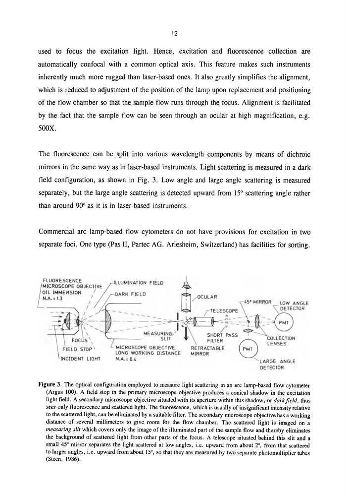
mirrors in the same way as in laser-based instruments. Light scattering is measured in a dark
field configuration, as shown in Fig. 3. Low angle and large angle scattering is measured
separately, but the large angle scattering is detected upward from 15° scattering angle rather
than around 90° as it is in laser-based instruments.
Commercial arc lamp-based flow cytometers do not have provisions for excitation in two
separate foci. One type (Pas II, Partec AG. Arlesheim, Switzerland) has facilities for sorting.
FLUORESCENCE
ILLUMINATION FIELD
~
t
MICROSCOPE OBJECTj/,VE
~
OIL IMMERSION
I jfDARK FIELD
m
N A-I 3
I
/ OCULAR
. . - .
I
' \45' MIRROR
lOW ANGLE
-,/// ~ --_____
I '
r TELES;OPE_ \ _ _ ~ \ DETECTOR
I",
- -
~
...,J I _ -
.....
\~:'~' ~~ ~-' -:E~s:~~)~t\ ~~{~:~- + > PMT
FOCUS \
SLIT
l FILTER e COLLECTION
\
LENSES
FIELD STOP \
MICROSCOPE OBJECTIVE
RETRACTABLE
PMT
LONG WORKING DISTANCE
MIRROR
INClOENT LIGHT
N.A.o 0.4
' LARGE ANGLE
DETECTOR
Figure 3. The optical configuration employed to measure light scattering in an arc lamp-based flow cytometer
(Argus 100). A field stop in the primary microscope objective produces a conical shadow in the excitation
light field. A secondary microscope objective situated with its aperture within this shadow, or darkfield, thus
sees only fluorescence and scattered light. The fluorescence, which is usually of insignificant intensity relative
to the scattered light, can be eliminated by a suitable filter. The secondary microscope objective has a working
distance of several millimeters to give room for the flow chamber. The scattered light is imaged on a
measuring slit which covers only the image of the illuminated part of the sample flow and thereby eliminates
the background of scattered light from other parts of the focus. A telescope situated behind this slit and a
small 45° mirror separates the light scattered at low angles, i.e. upward from about 2°, from that scattered
to larger angles, i.e. upward from about 15°, so that they are measured by two separate photomultiplier tubes
(Steen, 1986).
used to focus the excitation light. Hence, excitation and fluorescence collection are
automatically confocal with a common optical axis. This feature makes such instruments
inherently much more rugged than laser-based ones. It also greatly simplifies the alignment,
which is reduced to adjustment of the position of the lamp upon replacement and positioning
of the flow chamber so that the sample flow runs through the focus . Alignment is facilitated
by the fact that the sample flow can be seen through an ocular at high magnification, e.g.
500X.
The fluorescence can be split into various wavelength components by means of dichroic
mirrors in the same way as in laser-based instruments. Light scattering is measured in a dark
field configuration, as shown in Fig. 3. Low angle and large angle scattering is measured
separately, but the large angle scattering is detected upward from 15° scattering angle rather
than around 90° as it is in laser-based instruments.
Commercial arc lamp-based flow cytometers do not have provisions for excitation in two
separate foci. One type (Pas II, Partec AG. Arlesheim, Switzerland) has facilities for sorting.
FLUORESCENCE
ILLUMINATION FIELD
~
t
MICROSCOPE OBJECTj/,VE
~
OIL IMMERSION
I jfDARK FIELD
m
N A-I 3
I
/ OCULAR
. . - .
I
' \45' MIRROR
lOW ANGLE
-,/// ~ --_____
I '
r TELES;OPE_ \ _ _ ~ \ DETECTOR
I",
- -
~
...,J I _ -
.....
\~:'~' ~~ ~-' -:E~s:~~)~t\ ~~{~:~- + > PMT
FOCUS \
SLIT
l FILTER e COLLECTION
\
LENSES
FIELD STOP \
MICROSCOPE OBJECTIVE
RETRACTABLE
PMT
LONG WORKING DISTANCE
MIRROR
INClOENT LIGHT
N.A.o 0.4
' LARGE ANGLE
DETECTOR
Figure 3. The optical configuration employed to measure light scattering in an arc lamp-based flow cytometer
(Argus 100). A field stop in the primary microscope objective produces a conical shadow in the excitation
light field. A secondary microscope objective situated with its aperture within this shadow, or darkfield, thus
sees only fluorescence and scattered light. The fluorescence, which is usually of insignificant intensity relative
to the scattered light, can be eliminated by a suitable filter. The secondary microscope objective has a working
distance of several millimeters to give room for the flow chamber. The scattered light is imaged on a
measuring slit which covers only the image of the illuminated part of the sample flow and thereby eliminates
the background of scattered light from other parts of the focus. A telescope situated behind this slit and a
small 45° mirror separates the light scattered at low angles, i.e. upward from about 2°, from that scattered
to larger angles, i.e. upward from about 15°, so that they are measured by two separate photomultiplier tubes
(Steen, 1986).