

130
One problem has to do with differences in the optical properties of individual cells (Iturriaga
and Siegel, 1989). On the assumption that each particle is uniformly illuminated within the
flow cell and that fluorescence detection is equally uniform (independent of the transverse
position of the particle in the flow), then the amount of fluorescence (F) detected from each
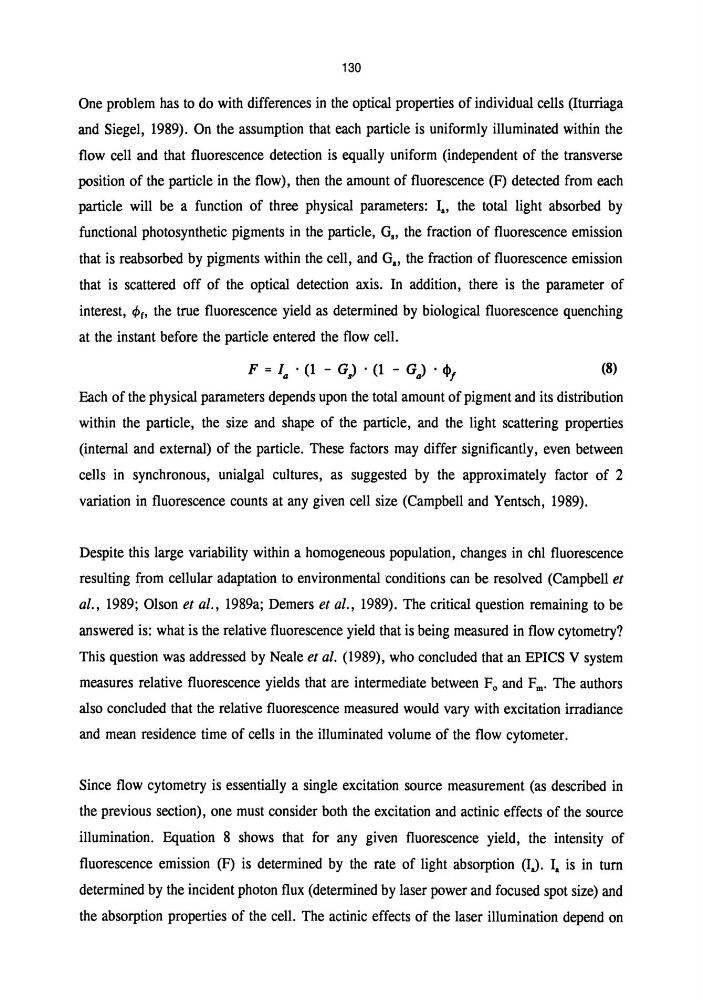
particle will be a function of three physical parameters: la, the total light absorbed by
functional photosynthetic pigments in the particle, G" the fraction of fluorescence emission
that is reabsorbed by pigments within the cell, and G .. the fraction of fluorescence emission
that is scattered off of the optical detection axis. In addition, there is the parameter of
interest, ¢f, the true fluorescence yield as determined by biological fluorescence quenching
at the instant before the particle entered the flow cell.
(8)
Each of the physical parameters depends upon the total amount of pigment and its distribution
within the particle, the size and shape of the particle, and the light scattering properties
(internal and external) of the particle. These factors may differ significantly, even between
cells in synchronous, unialgal cultures, as suggested by the approximately factor of 2
variation in fluorescence counts at any given cell size (Campbell and Yentsch, 1989).
Despite this large variability within a homogeneous population, changes in chI fluorescence
resulting from cellular adaptation to environmental conditions can be resolved (Campbell et
al., 1989; Olson et al., 1989a; Demers et al., 1989). The critical question remaining to be
answered is: what is the relative fluorescence yield that is being measured in flow cytometry?
This question was addressed by Neale et al. (1989), who concluded that an EPICS V system
measures relative fluorescence yields that are intermediate between F 0 and F m' The authors
also concluded that the relative fluorescence measured would vary with excitation irradiance
and mean residence time of cells in the illuminated volume of the flow cytometer.
Since flow cytometry is essentially a single excitation source measurement (as described in
the previous section), one must consider both the excitation and actinic effects of the source
illumination. Equation 8 shows that for any given fluorescence yield, the intensity of
fluorescence emission (F) is determined by the rate of light absorption (IJ. I. is in turn
determined by the incident photon flux (determined by laser power and focused spot size) and
the absorption properties of the cell. The actinic effects of the laser illumination depend on
One problem has to do with differences in the optical properties of individual cells (Iturriaga
and Siegel, 1989). On the assumption that each particle is uniformly illuminated within the
flow cell and that fluorescence detection is equally uniform (independent of the transverse
position of the particle in the flow), then the amount of fluorescence (F) detected from each
particle will be a function of three physical parameters: la, the total light absorbed by
functional photosynthetic pigments in the particle, G" the fraction of fluorescence emission
that is reabsorbed by pigments within the cell, and G .. the fraction of fluorescence emission
that is scattered off of the optical detection axis. In addition, there is the parameter of
interest, ¢f, the true fluorescence yield as determined by biological fluorescence quenching
at the instant before the particle entered the flow cell.
(8)
Each of the physical parameters depends upon the total amount of pigment and its distribution
within the particle, the size and shape of the particle, and the light scattering properties
(internal and external) of the particle. These factors may differ significantly, even between
cells in synchronous, unialgal cultures, as suggested by the approximately factor of 2
variation in fluorescence counts at any given cell size (Campbell and Yentsch, 1989).
Despite this large variability within a homogeneous population, changes in chI fluorescence
resulting from cellular adaptation to environmental conditions can be resolved (Campbell et
al., 1989; Olson et al., 1989a; Demers et al., 1989). The critical question remaining to be
answered is: what is the relative fluorescence yield that is being measured in flow cytometry?
This question was addressed by Neale et al. (1989), who concluded that an EPICS V system
measures relative fluorescence yields that are intermediate between F 0 and F m' The authors
also concluded that the relative fluorescence measured would vary with excitation irradiance
and mean residence time of cells in the illuminated volume of the flow cytometer.
Since flow cytometry is essentially a single excitation source measurement (as described in
the previous section), one must consider both the excitation and actinic effects of the source
illumination. Equation 8 shows that for any given fluorescence yield, the intensity of
fluorescence emission (F) is determined by the rate of light absorption (IJ. I. is in turn
determined by the incident photon flux (determined by laser power and focused spot size) and
the absorption properties of the cell. The actinic effects of the laser illumination depend on