

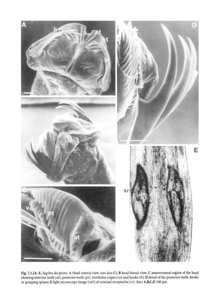
Fig. 7.1.2A-E. Sagitta decipiens. A Head ventral view (see also C); B head dorsal view; C anteroventral region of the head
showing anterior teeth (at), posterior teeth (pt), vestibular organ ( vo) and hooks (h); D detail of the posterior teeth, hooks
or grasping spines; E light microscope image (x45) of seminal receptacles (sr). Bars A,B,C,D 100 11m
showing anterior teeth (at), posterior teeth (pt), vestibular organ ( vo) and hooks (h); D detail of the posterior teeth, hooks
or grasping spines; E light microscope image (x45) of seminal receptacles (sr). Bars A,B,C,D 100 11m