

257
generation E� tasmanica females lay up to 5 clutches/lifetime, with sizes ranging
from 100 to 250 eggs per clutch. Hatching rate from the F2 clutches is approximately 99 % for the first clutch, with a decrease leading up to approximately 20 %
(~80 % hatching rate) for later clutches. F1 adults are larger and thus far have lived
for 1 year in captivity (Nishiguchi, unpublished). F2 adults have similar longevities,
but hatching rates for the F3 generation was somewhat lower (70–80 %)
15.4 Nursing of Eggs
15.4.1 Egg Characteristics
Eggs are single, stalkless and opaque white, having a droplet shape and calcareous
leather-like coating capsule (Fig. 15.3). The size of each egg is about 4 mm along
its major axis, 3 mm in its minor axis and weighs about 0.02 g. About 2 h after being laid, the outer coat (or capsule) turns brown, leathery and rigid in E� hyllebergi
(Nabhitabhata et al. 2005), whereas in E� tasmanica the egg is orange from wildcaught specimens. In F1 and subsequent generations of E� tasmanica, egg capsules
are white to opaque and remain so during development. This solid protection allows
the developing embryo to become a “sessile organism” during the extended period of development (Boletzky 1998). Eggs are telolecithal. Asymmetric eight-cell
cleavage occurs 10 h after fertilization. Clockwise rotation of the embryo occurs
from days 3 to 8, at 28 °C. Organogenesis occurs from day 4. The unique bilobed
character of the external yolk sac appears after day 5 when the capsule becomes
more transparent and the embryo is now visible. Chromatophores develop from day
6, and four diverticula of the internal yolk sac from day 8. The first hatching occurs
at day 12 (Fig. 15.4) for E� hyllebergi, and day 32 for E� tasmanica. The embryonic
phase is about 12–18 days, after approximately 14 days at 28 °C for E� hyllebergi,
and 21–28 days at 18 °C for E� tasmanica. The hatching period of eggs in the same
clutch takes 5 days from first to the last eggs and primarily occurs on the third day.
Average hatching rate is about 94 % (82–100 %) for both species.
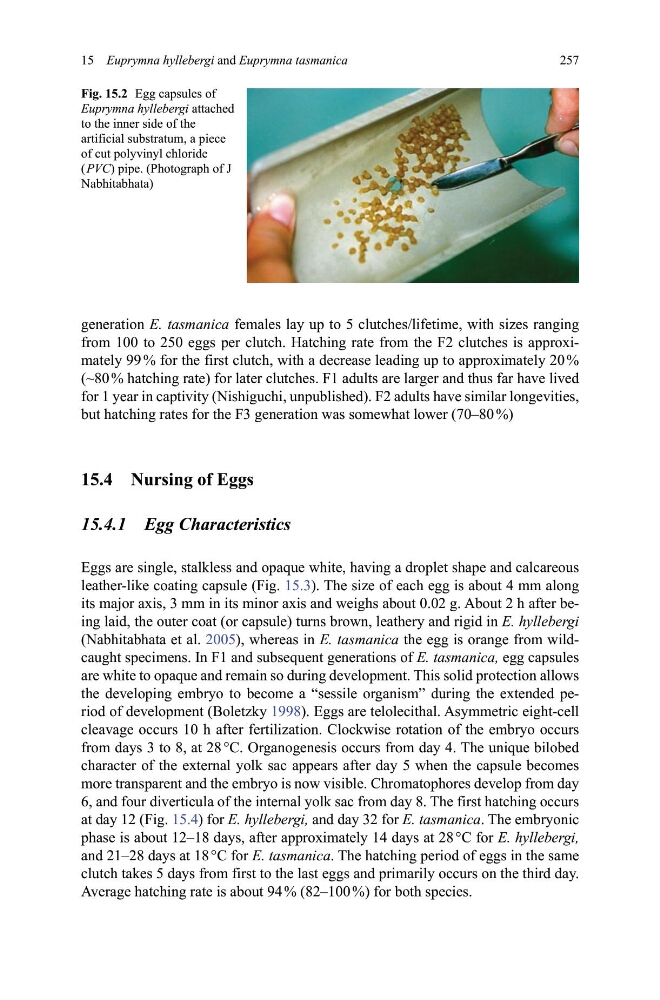
Fig. 15.2 Egg capsules of
Euprymna hyllebergi attached
to the inner side of the
artificial substratum, a piece
of cut polyvinyl chloride
( PVC) pipe. (Photograph of J
Nabhitabhata)
15 Euprymna hyllebergi and Euprymna tasmanica
generation E� tasmanica females lay up to 5 clutches/lifetime, with sizes ranging
from 100 to 250 eggs per clutch. Hatching rate from the F2 clutches is approximately 99 % for the first clutch, with a decrease leading up to approximately 20 %
(~80 % hatching rate) for later clutches. F1 adults are larger and thus far have lived
for 1 year in captivity (Nishiguchi, unpublished). F2 adults have similar longevities,
but hatching rates for the F3 generation was somewhat lower (70–80 %)
15.4 Nursing of Eggs
15.4.1 Egg Characteristics
Eggs are single, stalkless and opaque white, having a droplet shape and calcareous
leather-like coating capsule (Fig. 15.3). The size of each egg is about 4 mm along
its major axis, 3 mm in its minor axis and weighs about 0.02 g. About 2 h after being laid, the outer coat (or capsule) turns brown, leathery and rigid in E� hyllebergi
(Nabhitabhata et al. 2005), whereas in E� tasmanica the egg is orange from wildcaught specimens. In F1 and subsequent generations of E� tasmanica, egg capsules
are white to opaque and remain so during development. This solid protection allows
the developing embryo to become a “sessile organism” during the extended period of development (Boletzky 1998). Eggs are telolecithal. Asymmetric eight-cell
cleavage occurs 10 h after fertilization. Clockwise rotation of the embryo occurs
from days 3 to 8, at 28 °C. Organogenesis occurs from day 4. The unique bilobed
character of the external yolk sac appears after day 5 when the capsule becomes
more transparent and the embryo is now visible. Chromatophores develop from day
6, and four diverticula of the internal yolk sac from day 8. The first hatching occurs
at day 12 (Fig. 15.4) for E� hyllebergi, and day 32 for E� tasmanica. The embryonic
phase is about 12–18 days, after approximately 14 days at 28 °C for E� hyllebergi,
and 21–28 days at 18 °C for E� tasmanica. The hatching period of eggs in the same
clutch takes 5 days from first to the last eggs and primarily occurs on the third day.
Average hatching rate is about 94 % (82–100 %) for both species.
Fig. 15.2 Egg capsules of
Euprymna hyllebergi attached
to the inner side of the
artificial substratum, a piece
of cut polyvinyl chloride
( PVC) pipe. (Photograph of J
Nabhitabhata)
15 Euprymna hyllebergi and Euprymna tasmanica