

cytoarchitecture and connectivity, with a third para-belt region positioned
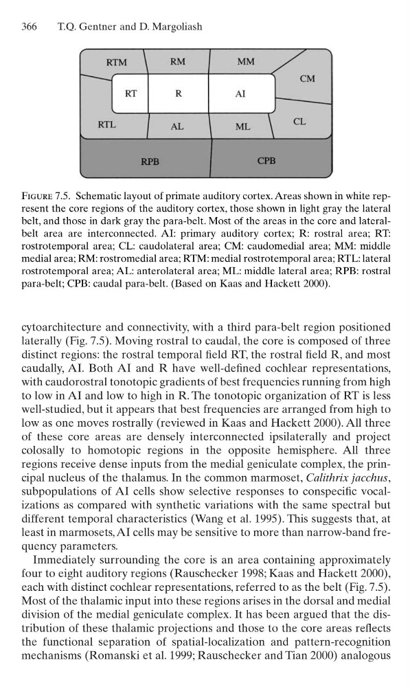
laterally (Fig. 7.5). Moving rostral to caudal, the core is composed of three
distinct regions: the rostral temporal field RT, the rostral field R, and most
caudally, AI. Both AI and R have well-defined cochlear representations,
with caudorostral tonotopic gradients of best frequencies running from high
to low in AI and low to high in R. The tonotopic organization of RT is less
well-studied, but it appears that best frequencies are arranged from high to
low as one moves rostrally (reviewed in Kaas and Hackett 2000). All three
of these core areas are densely interconnected ipsilaterally and project
colosally to homotopic regions in the opposite hemisphere. All three
regions receive dense inputs from the medial geniculate complex, the principal nucleus of the thalamus. In the common marmoset, Calithrix jacchus,
subpopulations of AI cells show selective responses to conspecific vocalizations as compared with synthetic variations with the same spectral but
different temporal characteristics (Wang et al. 1995). This suggests that, at
least in marmosets, AI cells may be sensitive to more than narrow-band frequency parameters.
Immediately surrounding the core is an area containing approximately
four to eight auditory regions (Rauschecker 1998; Kaas and Hackett 2000),
each with distinct cochlear representations, referred to as the belt (Fig. 7.5).
Most of the thalamic input into these regions arises in the dorsal and medial
division of the medial geniculate complex. It has been argued that the distribution of these thalamic projections and those to the core areas reflects
the functional separation of spatial-localization and pattern-recognition
mechanisms (Romanski et al. 1999; Rauschecker and Tian 2000) analogous
366
T.Q. Gentner and D. Margoliash
Figure 7.5. Schematic layout of primate auditory cortex. Areas shown in white represent the core regions of the auditory cortex, those shown in light gray the lateral
belt, and those in dark gray the para-belt. Most of the areas in the core and lateralbelt area are interconnected. AI: primary auditory cortex; R: rostral area; RT:
rostrotemporal area; CL: caudolateral area; CM: caudomedial area; MM: middle
medial area; RM: rostromedial area; RTM: medial rostrotemporal area; RTL: lateral
rostrotemporal area; AL: anterolateral area; ML: middle lateral area; RPB: rostral
para-belt; CPB: caudal para-belt. (Based on Kaas and Hackett 2000).
laterally (Fig. 7.5). Moving rostral to caudal, the core is composed of three
distinct regions: the rostral temporal field RT, the rostral field R, and most
caudally, AI. Both AI and R have well-defined cochlear representations,
with caudorostral tonotopic gradients of best frequencies running from high
to low in AI and low to high in R. The tonotopic organization of RT is less
well-studied, but it appears that best frequencies are arranged from high to
low as one moves rostrally (reviewed in Kaas and Hackett 2000). All three
of these core areas are densely interconnected ipsilaterally and project
colosally to homotopic regions in the opposite hemisphere. All three
regions receive dense inputs from the medial geniculate complex, the principal nucleus of the thalamus. In the common marmoset, Calithrix jacchus,
subpopulations of AI cells show selective responses to conspecific vocalizations as compared with synthetic variations with the same spectral but
different temporal characteristics (Wang et al. 1995). This suggests that, at
least in marmosets, AI cells may be sensitive to more than narrow-band frequency parameters.
Immediately surrounding the core is an area containing approximately
four to eight auditory regions (Rauschecker 1998; Kaas and Hackett 2000),
each with distinct cochlear representations, referred to as the belt (Fig. 7.5).
Most of the thalamic input into these regions arises in the dorsal and medial
division of the medial geniculate complex. It has been argued that the distribution of these thalamic projections and those to the core areas reflects
the functional separation of spatial-localization and pattern-recognition
mechanisms (Romanski et al. 1999; Rauschecker and Tian 2000) analogous
366
T.Q. Gentner and D. Margoliash
Figure 7.5. Schematic layout of primate auditory cortex. Areas shown in white represent the core regions of the auditory cortex, those shown in light gray the lateral
belt, and those in dark gray the para-belt. Most of the areas in the core and lateralbelt area are interconnected. AI: primary auditory cortex; R: rostral area; RT:
rostrotemporal area; CL: caudolateral area; CM: caudomedial area; MM: middle
medial area; RM: rostromedial area; RTM: medial rostrotemporal area; RTL: lateral
rostrotemporal area; AL: anterolateral area; ML: middle lateral area; RPB: rostral
para-belt; CPB: caudal para-belt. (Based on Kaas and Hackett 2000).