

beating wings, respectively. These multiple regions form a frequency versus
frequency topographical representation that systematically maps relative
target velocity. Another class of spectral combination-sensitive cells is
found in the Doppler-shift constant-frequency (DSCF) region of the cortex.
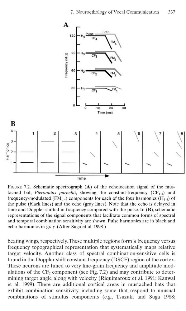
These neurons are tuned to very fine-grain frequency and amplitude modulations of the CF 2 component (see Fig. 7.2) and may contribute to determining target angle along with velocity (Riquimaroux et al. 1991; Kanwal
et al. 1999). There are additional cortical areas in mustached bats that
exhibit combination sensitivity, including some that respond to unusual
combinations of stimulus components (e.g., Tsuzuki and Suga 1988;
7. Neuroethology of Vocal Communication
337
4
3
2
1
Harmonics
Figure 7.2. Schematic spectrograph (A) of the echolocation signal of the mustached bat, Pteronutus parnellii, showing the constant-frequency (CF 1–4 ) and
frequency-modulated (FM 1–4 ) components for each of the four harmonics (H 1–4 ) of
the pulse (black lines) and the echo (gray lines). Note that the echo is delayed in
time and Doppler-shifted in frequency compared with the pulse. In (B), schematic
representations of the signal components that facilitate common forms of spectral
and temporal combination sensitivity are shown. Pulse harmonics are in black and
echo harmonics in gray. (After Suga et al. 1998.)
frequency topographical representation that systematically maps relative
target velocity. Another class of spectral combination-sensitive cells is
found in the Doppler-shift constant-frequency (DSCF) region of the cortex.
These neurons are tuned to very fine-grain frequency and amplitude modulations of the CF 2 component (see Fig. 7.2) and may contribute to determining target angle along with velocity (Riquimaroux et al. 1991; Kanwal
et al. 1999). There are additional cortical areas in mustached bats that
exhibit combination sensitivity, including some that respond to unusual
combinations of stimulus components (e.g., Tsuzuki and Suga 1988;
7. Neuroethology of Vocal Communication
337
4
3
2
1
Harmonics
Figure 7.2. Schematic spectrograph (A) of the echolocation signal of the mustached bat, Pteronutus parnellii, showing the constant-frequency (CF 1–4 ) and
frequency-modulated (FM 1–4 ) components for each of the four harmonics (H 1–4 ) of
the pulse (black lines) and the echo (gray lines). Note that the echo is delayed in
time and Doppler-shifted in frequency compared with the pulse. In (B), schematic
representations of the signal components that facilitate common forms of spectral
and temporal combination sensitivity are shown. Pulse harmonics are in black and
echo harmonics in gray. (After Suga et al. 1998.)