

10. Acoustic Models of Sound Production
419
Right Vestibular sac
Left
A.
Right arial
sac
sac
Right Premaxillary
ac
Left Vestibular
Right Vestibular ac
Right Premaxillary
"B.
sac
sac
arial
a
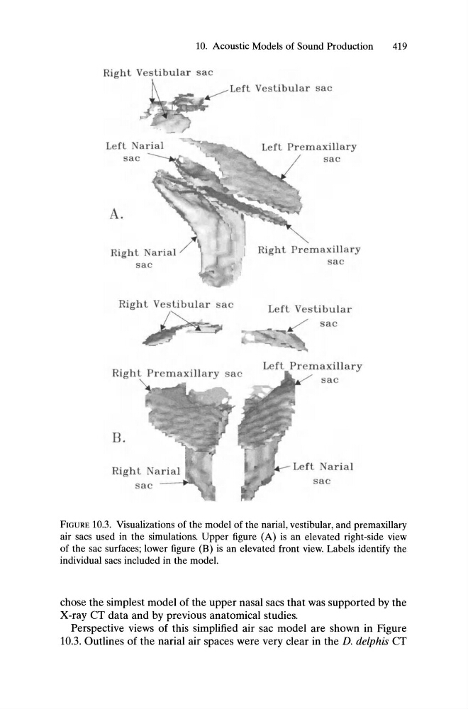
FIGURE 10.3. Visualizations of the model of the narial, vestibular, and premaxillary
air sacs used in the simulations. Upper figure (A) is an elevated right-side view
of the sac surfaces; lower figure (B) is an elevated front view. Labels identify the
individual sacs included in the model.
chose the simplest model of the upper nasal sacs that was supported by the
X-ray CT data and by previous anatomical studies.
Perspective views of this simplified air sac model are shown in Figure
10.3. Outlines of the narial air spaces were very clear in the D. de/phis CT
419
Right Vestibular sac
Left
A.
Right arial
sac
sac
Right Premaxillary
ac
Left Vestibular
Right Vestibular ac
Right Premaxillary
"B.
sac
sac
arial
a
FIGURE 10.3. Visualizations of the model of the narial, vestibular, and premaxillary
air sacs used in the simulations. Upper figure (A) is an elevated right-side view
of the sac surfaces; lower figure (B) is an elevated front view. Labels identify the
individual sacs included in the model.
chose the simplest model of the upper nasal sacs that was supported by the
X-ray CT data and by previous anatomical studies.
Perspective views of this simplified air sac model are shown in Figure
10.3. Outlines of the narial air spaces were very clear in the D. de/phis CT