

282
S.H. Ridgway
need such a large cerebellum. It has long been known that the cerebellum
has a great deal to do with fine motor movements and with the memory of
such movements. More recently, there has been considerable documentation of the involvement of the cerebellum in acoustic timing. For example,
cerebellar neurons function in circadian timing (Fauteck et al. 1994) and in
sound localization in the big brown bat, Eptesicus Juscus, (Kamada and Jen
1990) and in the acoustics of the biosonar signals and frequency sensitivity
in the mustached bat Pteronotus parnelli (Horikawa and Suga 1986). The
involvement of the cerebellum in acoustic processing would seem to help
explain why delphinoids have such a large cerebellum.
5. The Auditory Cortex
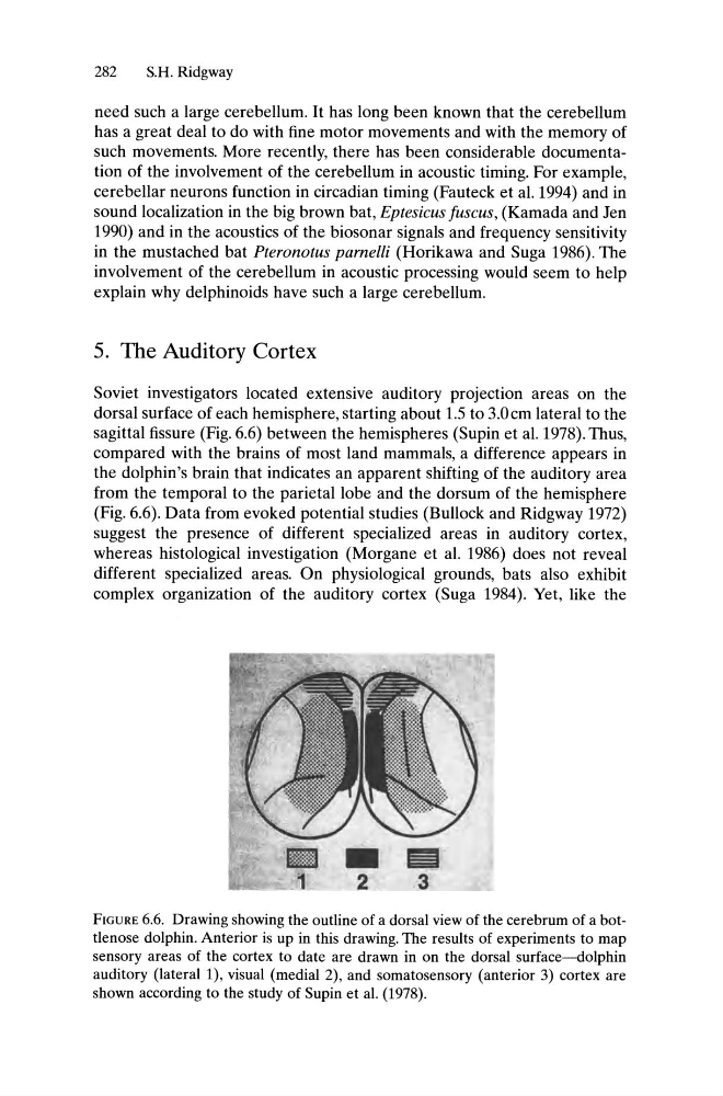
Soviet investigators located extensive auditory projection areas on the
dorsal surface of each hemisphere, starting about 1.5 to 3.0cm lateral to the
sagittal fissure (Fig. 6.6) between the hemispheres (Supin et al. 1978). Thus,
compared with the brains of most land mammals, a difference appears in
the dolphin's brain that indicates an apparent shifting of the auditory area
from the temporal to the parietal lobe and the dorsum of the hemisphere
(Fig. 6.6). Data from evoked potential studies (Bullock and Ridgway 1972)
suggest the presence of different specialized areas in auditory cortex,
whereas histological investigation (Morgane et al. 1986) does not reveal
different specialized areas. On physiological grounds, bats also exhibit
complex organization of the auditory cortex (Suga 1984). Yet, like the
FIGURE 6.6. Drawing showing the outline of a dorsal view of the cerebrum of a bottlenose dolphin. Anterior is up in this drawing. The results of experiments to map
sensory areas of the cortex to date are drawn in on the dorsal surface~olphin
auditory (lateral 1), visual (medial 2), and somatosensory (anterior 3) cortex are
shown according to the study of Supin et al. (1978).
S.H. Ridgway
need such a large cerebellum. It has long been known that the cerebellum
has a great deal to do with fine motor movements and with the memory of
such movements. More recently, there has been considerable documentation of the involvement of the cerebellum in acoustic timing. For example,
cerebellar neurons function in circadian timing (Fauteck et al. 1994) and in
sound localization in the big brown bat, Eptesicus Juscus, (Kamada and Jen
1990) and in the acoustics of the biosonar signals and frequency sensitivity
in the mustached bat Pteronotus parnelli (Horikawa and Suga 1986). The
involvement of the cerebellum in acoustic processing would seem to help
explain why delphinoids have such a large cerebellum.
5. The Auditory Cortex
Soviet investigators located extensive auditory projection areas on the
dorsal surface of each hemisphere, starting about 1.5 to 3.0cm lateral to the
sagittal fissure (Fig. 6.6) between the hemispheres (Supin et al. 1978). Thus,
compared with the brains of most land mammals, a difference appears in
the dolphin's brain that indicates an apparent shifting of the auditory area
from the temporal to the parietal lobe and the dorsum of the hemisphere
(Fig. 6.6). Data from evoked potential studies (Bullock and Ridgway 1972)
suggest the presence of different specialized areas in auditory cortex,
whereas histological investigation (Morgane et al. 1986) does not reveal
different specialized areas. On physiological grounds, bats also exhibit
complex organization of the auditory cortex (Suga 1984). Yet, like the
FIGURE 6.6. Drawing showing the outline of a dorsal view of the cerebrum of a bottlenose dolphin. Anterior is up in this drawing. The results of experiments to map
sensory areas of the cortex to date are drawn in on the dorsal surface~olphin
auditory (lateral 1), visual (medial 2), and somatosensory (anterior 3) cortex are
shown according to the study of Supin et al. (1978).