

278
S.H. Ridgway
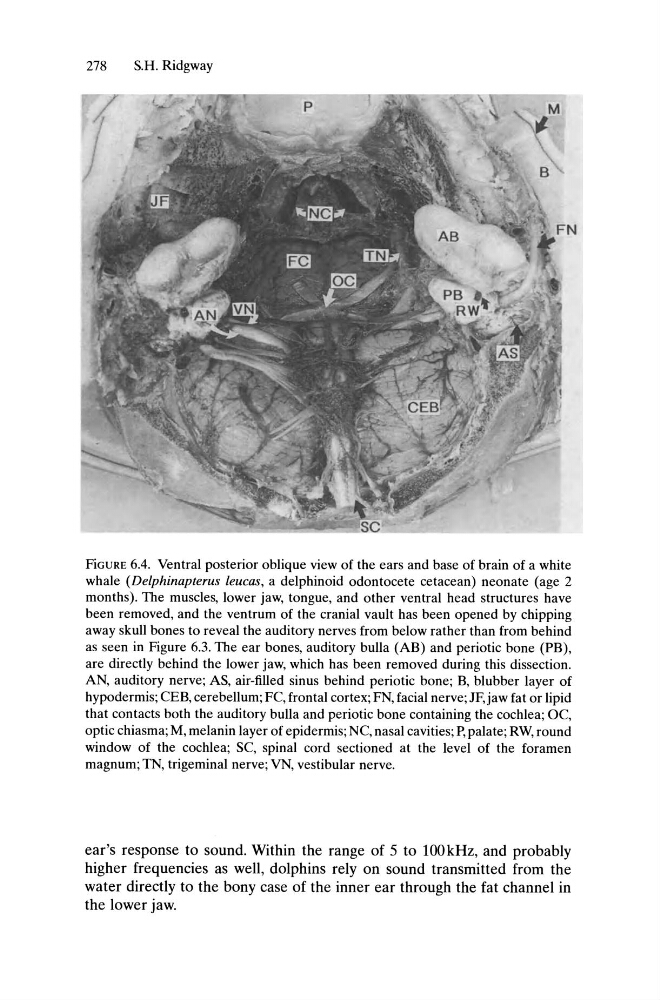
FiGURE 6.4. Ventral posterior oblique view of the ears and base of brain of a white
whale (Delphinapterus leucas, a delphinoid odontocete cetacean) neonate (age 2
months). The muscles, lower jaw, tongue, and other ventral head structures have
been removed, and the ventrum of the cranial vault has been opened by chipping
away skull bones to reveal the auditory nerves from below rather than from behind
as seen in Figure 6.3. The ear bones, auditory bulla (AB) and periotic bone (PB),
are directly behind the lower jaw, which has been removed during this dissection.
AN, auditory nerve; AS, air-filled sinus behind periotic bone; B, blubber layer of
hypodermis; CEB, cerebellum; FC, frontal cortex; FN, facial nerve; JF,jaw fat or lipid
that contacts both the auditory bulla and periotic bone containing the cochlea; OC,
optic chiasma; M, melanin layer of epidermis; NC, nasal cavities; P, palate; RW, round
window of the cochlea; SC, spinal cord sectioned at the level of the foramen
magnum; TN, trigeminal nerve; VN, vestibular nerve.
ear's response to sound. Within the range of 5 to 100 kHz, and probably
higher frequencies as well, dolphins rely on sound transmitted from the
water directly to the bony case of the inner ear through the fat channel in
the lower jaw.
S.H. Ridgway
FiGURE 6.4. Ventral posterior oblique view of the ears and base of brain of a white
whale (Delphinapterus leucas, a delphinoid odontocete cetacean) neonate (age 2
months). The muscles, lower jaw, tongue, and other ventral head structures have
been removed, and the ventrum of the cranial vault has been opened by chipping
away skull bones to reveal the auditory nerves from below rather than from behind
as seen in Figure 6.3. The ear bones, auditory bulla (AB) and periotic bone (PB),
are directly behind the lower jaw, which has been removed during this dissection.
AN, auditory nerve; AS, air-filled sinus behind periotic bone; B, blubber layer of
hypodermis; CEB, cerebellum; FC, frontal cortex; FN, facial nerve; JF,jaw fat or lipid
that contacts both the auditory bulla and periotic bone containing the cochlea; OC,
optic chiasma; M, melanin layer of epidermis; NC, nasal cavities; P, palate; RW, round
window of the cochlea; SC, spinal cord sectioned at the level of the foramen
magnum; TN, trigeminal nerve; VN, vestibular nerve.
ear's response to sound. Within the range of 5 to 100 kHz, and probably
higher frequencies as well, dolphins rely on sound transmitted from the
water directly to the bony case of the inner ear through the fat channel in
the lower jaw.