

90
D.R. Ketten
MYSTICETE
M
Type I
v
ODONTOCETE
v
Type II
p
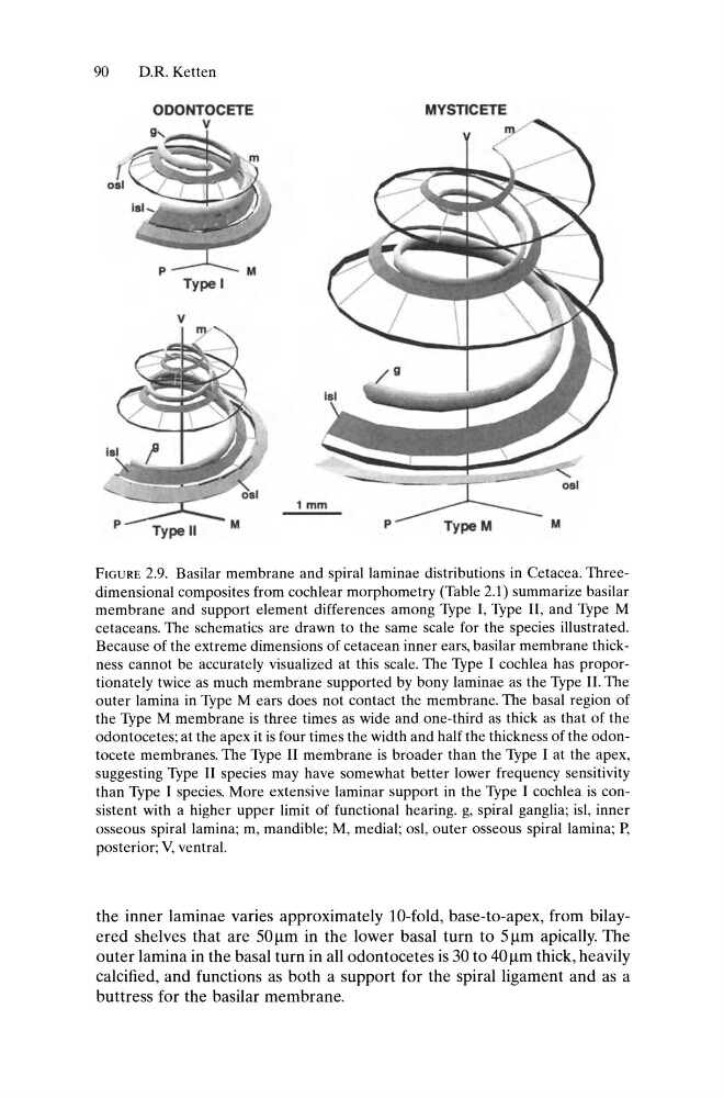
FIGURE 2.9. Basilar membrane and spiral laminae distributions in Cetacea. Threedimensional composites from cochlear morphometry (Table 2.1) summarize basilar
membrane and support element differences among Type I, Type II, and Type M
cetaceans. The schematics are drawn to the same scale for the species illustrated.
Because of the extreme dimensions of cetacean inner ears, basilar membrane thickness cannot be accurately visualized at this scale. The Type I cochlea has proportionately twice as much membrane supported by bony laminae as the Type II. The
outer lamina in Type M ears does not contact the membrane. The basal region of
the Type M membrane is three times as wide and one-third as thick as that of the
odontocetes; at the apex it is four times the width and half the thickness of the odontocete membranes. The Type II membrane is broader than the Type I at the apex,
suggesting Type II species may have somewhat better lower frequency sensitivity
than Type I species. More extensive laminar support in the Type I cochlea is consistent with a higher upper limit of functional hearing. g, spiral ganglia; isl, inner
osseous spiral lamina; m, mandible; M, medial; osl, outer osseous spiral lamina; P,
posterior; V, ventral.
the inner laminae varies approximately lO-fold, base-to-apex, from bilayered shelves that are 50 Ilm in the lower basal turn to 51lm apically. The
outer lamina in the basal turn in all odontocetes is 30 to 40 Ilm thick, heavily
calcified, and functions as both a support for the spiral ligament and as a
buttress for the basilar membrane.
D.R. Ketten
MYSTICETE
M
Type I
v
ODONTOCETE
v
Type II
p
FIGURE 2.9. Basilar membrane and spiral laminae distributions in Cetacea. Threedimensional composites from cochlear morphometry (Table 2.1) summarize basilar
membrane and support element differences among Type I, Type II, and Type M
cetaceans. The schematics are drawn to the same scale for the species illustrated.
Because of the extreme dimensions of cetacean inner ears, basilar membrane thickness cannot be accurately visualized at this scale. The Type I cochlea has proportionately twice as much membrane supported by bony laminae as the Type II. The
outer lamina in Type M ears does not contact the membrane. The basal region of
the Type M membrane is three times as wide and one-third as thick as that of the
odontocetes; at the apex it is four times the width and half the thickness of the odontocete membranes. The Type II membrane is broader than the Type I at the apex,
suggesting Type II species may have somewhat better lower frequency sensitivity
than Type I species. More extensive laminar support in the Type I cochlea is consistent with a higher upper limit of functional hearing. g, spiral ganglia; isl, inner
osseous spiral lamina; m, mandible; M, medial; osl, outer osseous spiral lamina; P,
posterior; V, ventral.
the inner laminae varies approximately lO-fold, base-to-apex, from bilayered shelves that are 50 Ilm in the lower basal turn to 51lm apically. The
outer lamina in the basal turn in all odontocetes is 30 to 40 Ilm thick, heavily
calcified, and functions as both a support for the spiral ligament and as a
buttress for the basilar membrane.