

465
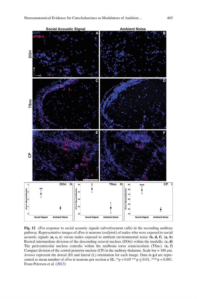
Fig. 12 cFos response to social acoustic signals (advertisement calls) in the ascending auditory
pathway. Representative images of cFos-ir neurons ( red/pink ) of males who were exposed to social
acoustic signals ( a, c, e ) versus males exposed to ambient environmental noise ( b, d, f ). ( a, b )
Rostral intermediate division of the descending octaval nucleus (DOri) within the medulla. ( c, d )
The periventricular nucleus centralis within the midbrain torus semicircularis (TSnc). ( e, f )
Compact division of the central posterior nucleus (CP) in the auditory thalamus. Scale bar = 100 μm.
Arrows represent the dorsal (D) and lateral (L) orientation for each image. Data in g-i are represented as mean number of cFos-ir neurons per section ± SE, * p < 0.05 ** p ≤ 0.01, *** p < 0.001.
From Petersen et al. ( 2013 )
Neuroanatomical Evidence for Catecholamines as Modulators of Audition…
Fig. 12 cFos response to social acoustic signals (advertisement calls) in the ascending auditory
pathway. Representative images of cFos-ir neurons ( red/pink ) of males who were exposed to social
acoustic signals ( a, c, e ) versus males exposed to ambient environmental noise ( b, d, f ). ( a, b )
Rostral intermediate division of the descending octaval nucleus (DOri) within the medulla. ( c, d )
The periventricular nucleus centralis within the midbrain torus semicircularis (TSnc). ( e, f )
Compact division of the central posterior nucleus (CP) in the auditory thalamus. Scale bar = 100 μm.
Arrows represent the dorsal (D) and lateral (L) orientation for each image. Data in g-i are represented as mean number of cFos-ir neurons per section ± SE, * p < 0.05 ** p ≤ 0.01, *** p < 0.001.
From Petersen et al. ( 2013 )
Neuroanatomical Evidence for Catecholamines as Modulators of Audition…