

458
and Panula 2001 ; Ma 1994 ; O’Connell et al. 2011 ; Roberts et al. 1989 ; Sas et al.
1990 ; Vetillard et al. 2002 ). In addition, several studies in teleosts have demonstrated DAergic and/or NAergic receptor distribution in auditory nuclei which supports the auditory system as a CA target for sensory modulation. Vacher et al. ( 2003 )
showed D 2 mRNA in TS (centralis and lateralis) in trout, and O’Connell et al. ( 2011 )
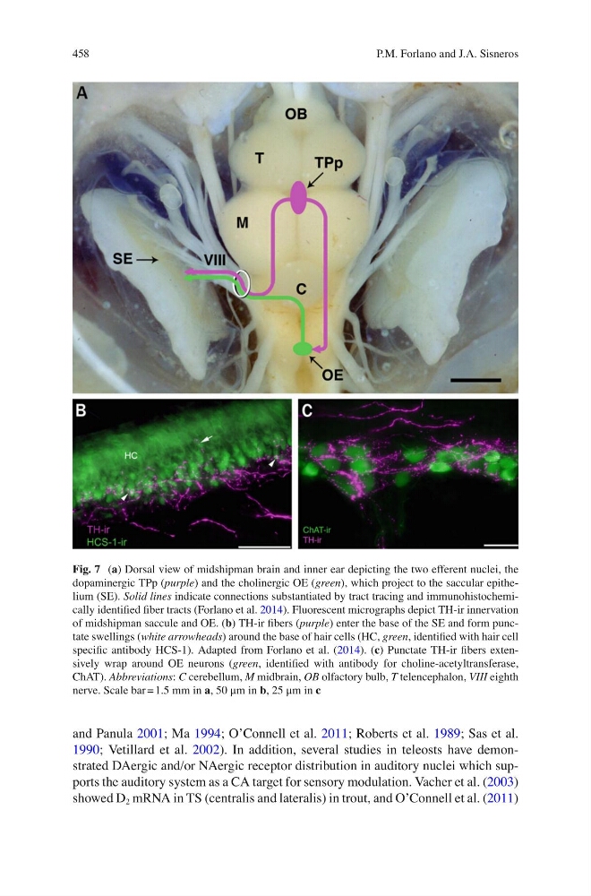
Fig. 7 ( a ) Dorsal view of midshipman brain and inner ear depicting the two efferent nuclei, the
dopaminergic TPp ( purple ) and the cholinergic OE ( green ), which project to the saccular epithelium (SE). Solid lines indicate connections substantiated by tract tracing and immunohistochemically identifi ed fi ber tracts (Forlano et al. 2014 ). Fluorescent micrographs depict TH-ir innervation
of midshipman saccule and OE. ( b ) TH-ir fi bers ( purple ) enter the base of the SE and form punctate swellings ( white arrowheads ) around the base of hair cells (HC, green , identifi ed with hair cell
specifi c antibody HCS-1). Adapted from Forlano et al. ( 2014 ). ( c ) Punctate TH-ir fi bers extensively wrap around OE neurons ( green , identifi ed with antibody for choline-acetyltransferase,
ChAT). Abbreviations : C cerebellum, M midbrain, OB olfactory bulb, T telencephalon, VIII eighth
nerve. Scale bar = 1.5 mm in a , 50 μm in b , 25 μm in c
P.M. Forlano and J.A. Sisneros
and Panula 2001 ; Ma 1994 ; O’Connell et al. 2011 ; Roberts et al. 1989 ; Sas et al.
1990 ; Vetillard et al. 2002 ). In addition, several studies in teleosts have demonstrated DAergic and/or NAergic receptor distribution in auditory nuclei which supports the auditory system as a CA target for sensory modulation. Vacher et al. ( 2003 )
showed D 2 mRNA in TS (centralis and lateralis) in trout, and O’Connell et al. ( 2011 )
Fig. 7 ( a ) Dorsal view of midshipman brain and inner ear depicting the two efferent nuclei, the
dopaminergic TPp ( purple ) and the cholinergic OE ( green ), which project to the saccular epithelium (SE). Solid lines indicate connections substantiated by tract tracing and immunohistochemically identifi ed fi ber tracts (Forlano et al. 2014 ). Fluorescent micrographs depict TH-ir innervation
of midshipman saccule and OE. ( b ) TH-ir fi bers ( purple ) enter the base of the SE and form punctate swellings ( white arrowheads ) around the base of hair cells (HC, green , identifi ed with hair cell
specifi c antibody HCS-1). Adapted from Forlano et al. ( 2014 ). ( c ) Punctate TH-ir fi bers extensively wrap around OE neurons ( green , identifi ed with antibody for choline-acetyltransferase,
ChAT). Abbreviations : C cerebellum, M midbrain, OB olfactory bulb, T telencephalon, VIII eighth
nerve. Scale bar = 1.5 mm in a , 50 μm in b , 25 μm in c
P.M. Forlano and J.A. Sisneros